You're viewing 2 free preview questions for Biology HL — subscribe to unlock 202 more, plus the Full Question Bank, Mock Papers & Worksheets.Upgrade
Proteins · Paper 2 — Free Biology HL Practice Questions
1MasterySAQ-SEnzymes as biological catalysts5 marksPaper 2~8 min

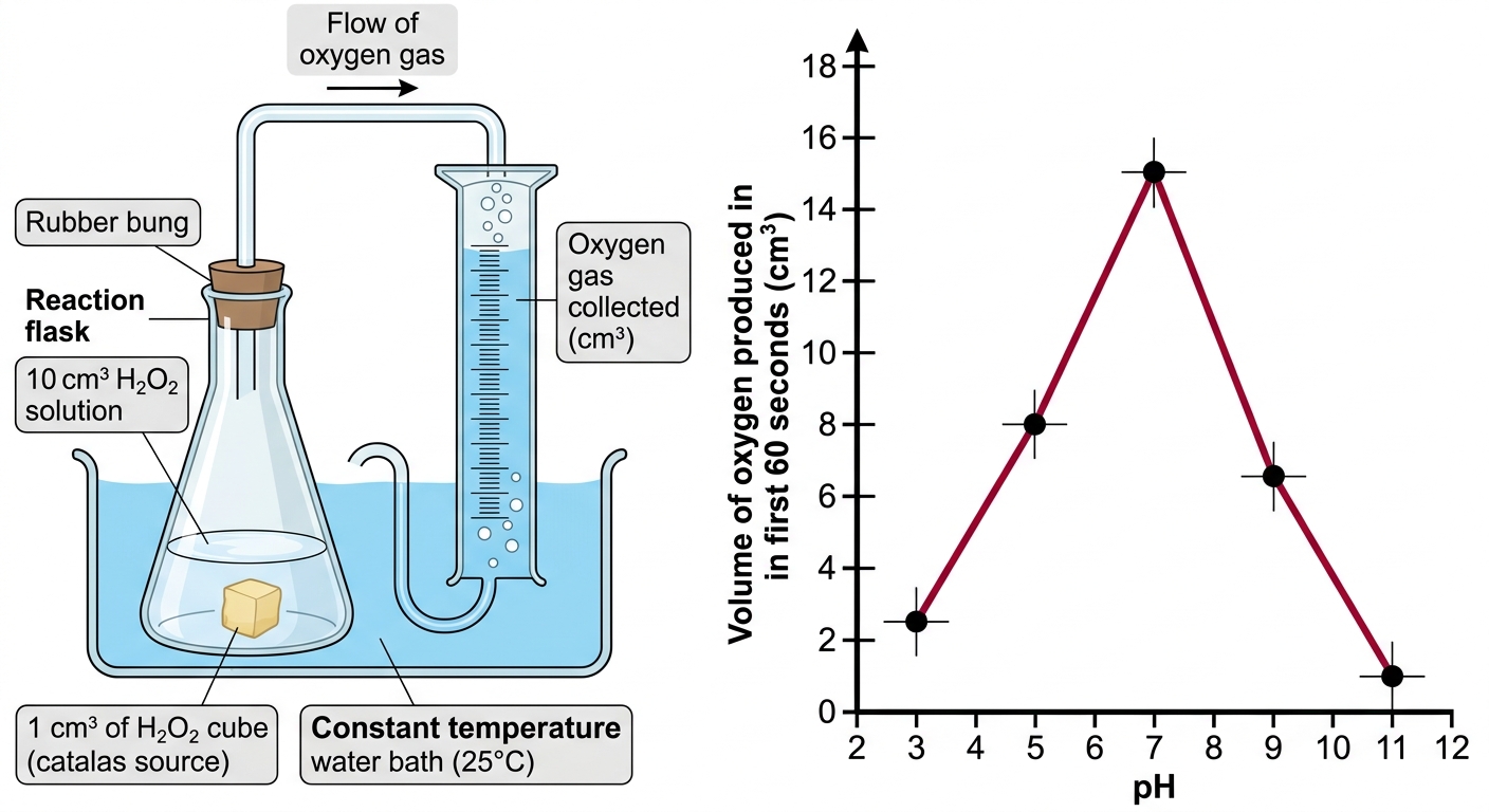
A student investigates the effect of pH on the activity of the enzyme catalase, which breaks down hydrogen peroxide () into water and oxygen. A cube of potato (a source of catalase) is added to of hydrogen peroxide solution at five different pH values. The volume of oxygen gas produced in the first 60 seconds is recorded.
pH — Volume of produced /
3 — 2.5
5 — 8.0
7 — 15.0
9 — 6.5
11 — 1.0
All experiments are conducted at .
(a)
State the pH at which catalase activity is highest. [1 mark]
(b)
Describe the effect of pH on the volume of oxygen produced, using the data provided. [2 marks]
(c)
Explain why catalase activity is lower at pH 3 than at pH 7. [2 marks]

Solutions
2ChallengeSAQ-LStructure of proteins: Primary to quaternary structure9 marksPaper 2~14 min
Data
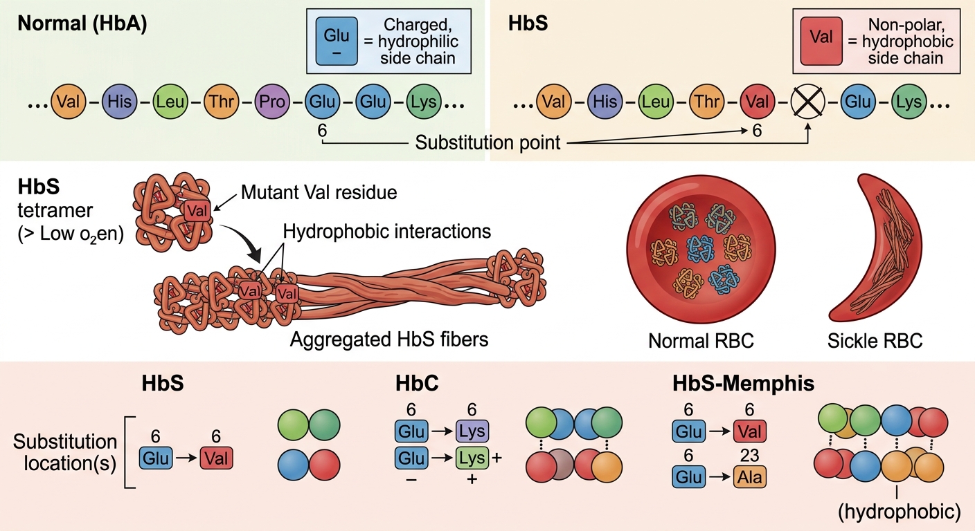
- Glutamic acid has a charged, hydrophilic R-group; valine has a non-polar, hydrophobic R-group.
- HbA is highly soluble in the cytoplasm of red blood cells and exists a tetramer () with quaternary structure.
- Under low-oxygen conditions, HbS molecules aggregate into long fibrous polymers that deform red blood cells into a sickle shape.
- A second mutant, HbC, has Glu at position 6 replaced by lysine (Lys), which has a positively charged R-group. HbC does not form polymers under low-oxygen conditions.
- A third mutant, HbS-Memphis, carries the same GluVal substitution at position 6 as HbS, plus a second substitution at position 23 where glutamic acid is replaced by alanine (Ala), which is hydrophobic. HbS-Memphis forms polymers more readily than HbS under the same conditions.
A molecular biology laboratory is studying a mutant form of haemoglobin, HbS, which causes sickle cell anaemia. Normal adult haemoglobin (HbA) has the following primary structure at the beginning of its beta-globin chain:
In HbS, a single nucleotide substitution in the beta-globin gene causes glutamic acid (Glu) at position 6 to be replaced by valine (Val).
Additional
(a)
State the level of protein structure directly altered by the GluVal substitution, and identify two higher levels of protein structure that are consequently affected in HbS. [2 marks]
(b)
Explain how the single GluVal substitution leads to polymer formation under low-oxygen conditions. [3 marks]
(c)
Evaluate the statement: "The quaternary structure of a protein is entirely determined by its primary structure," using evidence from at least three of the haemoglobin variants described above. [4 marks]
Solutions
That's every free question here — 2 of 204.
+202 more questions, plus Mock Papers & Worksheets, are waiting in Premium.