You're viewing 3 free preview questions for Biology SL — subscribe to unlock 45 more, plus the Full Question Bank, Mock Papers & Worksheets.Upgrade
Cell and Nuclear Division · Paper 2 — Free Biology SL Practice Questions
1MasterySAQ-SMitosis vs meiosis6 marksPaper 2~9 min

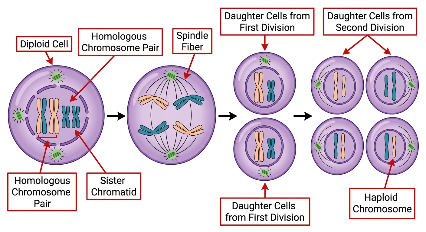
A single diploid cell ($2n = 4$) undergoes nuclear division as shown below.
Cell 1: Four chromosomes (two long, two short), each consisting of two sister chromatids, arranged as two homologous pairs.
Cell 2: Homologous pairs have separated; one long and one short chromosome move to opposite poles.
Cell 3: Two cells, each containing two chromosomes (one long, one short), each still consisting of two sister chromatids.
Cell 4: Four cells, each containing two chromosomes (one long, one short) as single chromatids.
(a)
State the type of nuclear division shown. [1 mark]
(b)
State the number of chromatids per cell at the stage shown in Cell 3, and explain why this differs from the number of chromosomes per cell at Cell 4. [2 marks]
(c)
Describe one event occurring during prophase I that increases genetic variation. [1 mark]
(d)
Explain how independent assortment, acting alone, produces genetic differences between the cells in Cell 4 and the original cell in Cell 1. [2 marks]

Solutions
2MasterySAQ-SPhases of mitosis and meiosis5 marksPaper 2~8 min
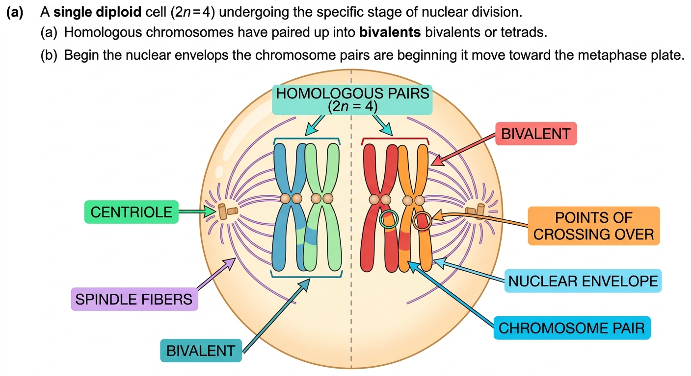
A diploid cell ($2n = 4$) undergoes meiosis. The shows the cell at metaphase I, with two homologous pairs of chromosomes arranged at the equator of the cell. Chiasmata are visible between homologous chromosomes.
(a)
State the stage of meiosis shown in the . [1 mark]
(b)
Describe the behaviour of chromosomes during metaphase I of meiosis. [2 marks]
(c)
A student examines 250 cells from anther undergoing meiosis and records that 20 cells are at metaphase I. The entire meiotic division takes 48 hours. Calculate the average time, in minutes, that a cell spends in metaphase I. Show your working. [2 marks]
Solutions
3ChallengeSAQ-LMitosis vs meiosis7 marksPaper 2~11 min
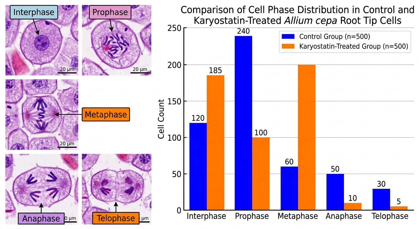
A research team investigates the effects of a novel compound, Karyostatin, on cell division in the root meristem of Allium cepa. Root tip cells are treated with Karyostatin for 24 hours, then fixed, stained, and observed under a light microscope. Cells are classified by phase. Results are shown below.
Phase — Control (500 cells) — Karyostatin-treated (500 cells)
Interphase — 120 — 185
Prophase — 240 — 100
Metaphase — 60 — 200
Anaphase — 50 — 10
Telophase — 30 — 5
(a)
Calculate the mitotic index for both the control group and the Karyostatin-treated group, expressing each as a percentage. [2 marks]
(b)
Using the data, explain the stage at which Karyostatin disrupts mitosis and identify the most likely cellular mechanism responsible. [3 marks]
(c)
Karyostatin is proposed as a cancer therapeutic. Evaluate the significance of the observed decrease in mitotic index in the treated group when assessing Karyostatin's potential effectiveness against rapidly dividing tumour cells. [2 marks]
Solutions
That's every free question here — 3 of 48.
+45 more questions, plus Mock Papers & Worksheets, are waiting in Premium.