Microscopes and Cell Observation — Free MYP1 Sciences Practice Questions

1QuestionParts and Functions of a Light MicroscopeConcept Practice
2 marks~3 minCriterion A
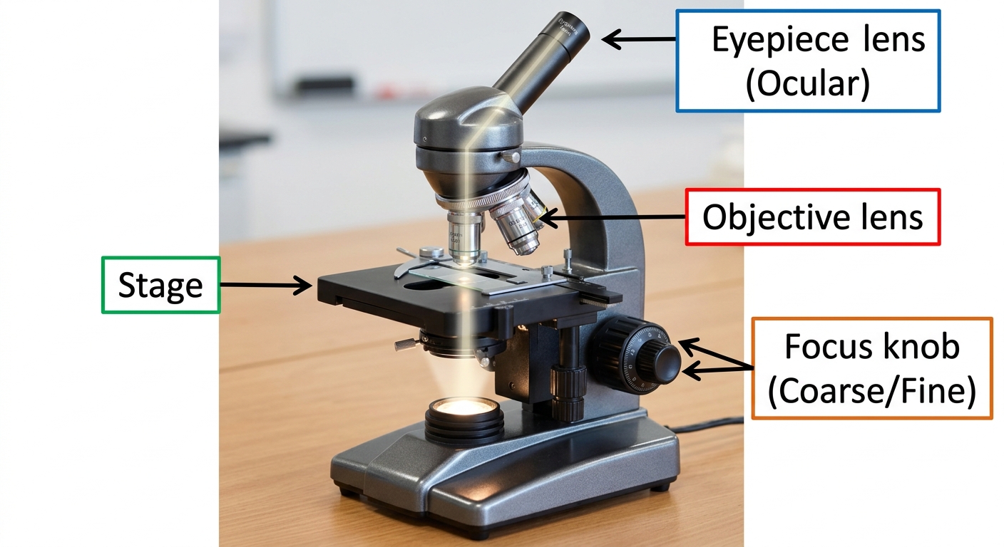
A light microscope has several important parts.
a
Name one job of the eyepiece lens. [1]
b
Describe how the stage and the focus knob each help you to observe a specimen. [1]

Solutions
2QuestionMaking Temporary Slides Onion Cells etcAssessment Practice
4 marks~6 minCriterion A
A student looked at onion cells under a microscope. The table below shows how many graticule divisions one cell measured at each lens setting.
Each graticule division equals 0.025 mm when the objective lens is used.
| Objective lens | |||
|---|---|---|---|
| Graticule divisions | 20 | 8 | ? |
Each graticule division equals 0.025 mm when the objective lens is used.
a
Identify the missing number of graticule divisions for the objective lens. [1]
b
State the actual length of the onion cell in mm. Show your working. [2]
c
Describe one way the image of the cell would look different when you switch from the lens to the lens. [1]
Solutions
3QuestionParts and Functions of a Light MicroscopeConcept Practice
2 marks~3 minCriterion A
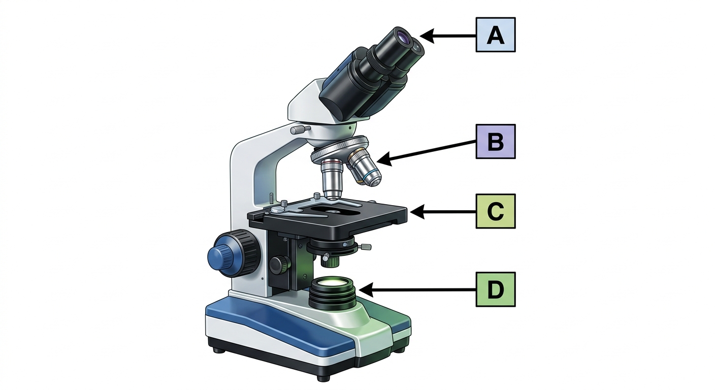
The diagram below shows a light microscope with four parts labelled A, B, C, and D.
A: Eyepiece
B: Objective lens
C: Stage
D: Light source
A: Eyepiece
B: Objective lens
C: Stage
D: Light source
a
Identify the letter that shows the part you look through to see a magnified specimen. [1]
b
Describe what the stage (C) is used for when looking at a specimen under the microscope. [1]
Solutions
4QuestionSafety and Handling of MicroscopesAssessment Practice
4 marks~6 minCriterion D
A school has 10 microscopes shared by 30 students. Each microscope costs 200 dollars to repair.
a
Identify one way a student could damage a microscope lens. [1]
b
Describe one effect on students' learning if a microscope is sent away for repair. [1]
c
A student says, "Carrying a microscope with two hands is annoying — it doesn't matter." Describe one reason why using two hands does matter, and explain how it affects both the school's money and students' learning time. [2]
Solutions
5QuestionParts and Functions of a Light MicroscopeConcept Practice
2 marks~3 minCriterion A
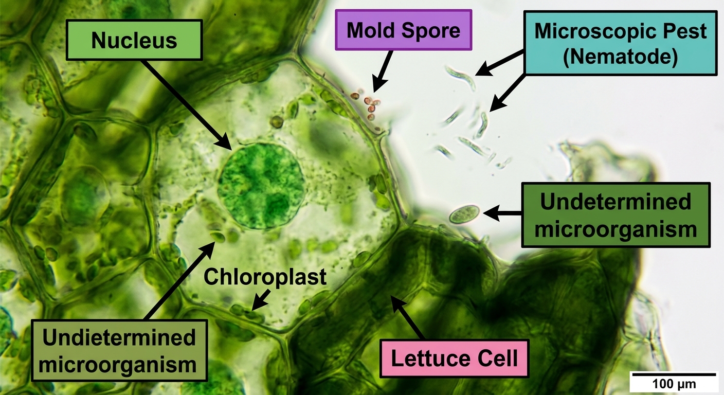
A food inspector uses a light microscope to check a lettuce sample for mold spores and tiny pests that cannot be seen with the naked eye.
a
State one way a light microscope helps keep food safe. [1]
b
Identify one thing a light microscope cannot do when checking food. [1]
Solutions
6QuestionParts and Functions of a Light MicroscopeAssessment Practice
5 marks~8 minCriterion A
A microscope is used to look at four different cells. The table below shows information about each image.
Image A: scale bar actual size = 10 µm, scale bar length on diagram = 20 mm
Image B: scale bar actual size = 50 µm, scale bar length on diagram = 25 mm
Image C: scale bar actual size = 5 µm, scale bar length on diagram = 15 mm
Image D: scale bar actual size = 100 µm, scale bar length on diagram = 10 mm
Use the formula:
Image A: scale bar actual size = 10 µm, scale bar length on diagram = 20 mm
Image B: scale bar actual size = 50 µm, scale bar length on diagram = 25 mm
Image C: scale bar actual size = 5 µm, scale bar length on diagram = 15 mm
Image D: scale bar actual size = 100 µm, scale bar length on diagram = 10 mm
Use the formula:
a
Name the part of a microscope that makes the image larger. [1]
b
Calculate the magnification for Image A. Show your working. [2]
c
Image C has a magnification of ×3000 and Image D has a magnification of ×100. Describe what these two numbers tell you about how the two images look different on the page. [2]
Solutions
7QuestionMaking Temporary Slides Onion Cells etcConcept Practice
2 marks~3 minCriterion A
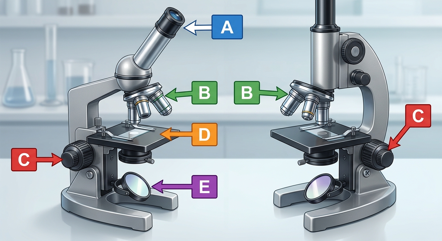
The diagram shows a compound light microscope with parts labeled A to E.
a
Identify the part labeled D. [1]
b
Describe what part D is used for when looking at an onion cell slide. [1]
Solutions
8QuestionUsing a Microscope to View Prepared SlidesAssessment Practice
5 marks~8 minCriterion A
A student looks at onion cells through a school microscope. On the low-power setting, the cells look clear. When the student switches to the high-power setting, the image becomes blurry and dark.
a
Name the part of the microscope the student changes when switching from low power to high power. [1]
b
Describe why the image becomes blurry when the student increases the magnification. [2]
c
The student wants to see ribosomes inside the cells. Ribosomes are much smaller than cell walls. Describe one difference between a school microscope and an electron microscope that explains why a school microscope cannot be used to see ribosomes. [2]
Solutions
9QuestionMagnification and Field of ViewConcept Practice
2 marks~3 minCriterion A
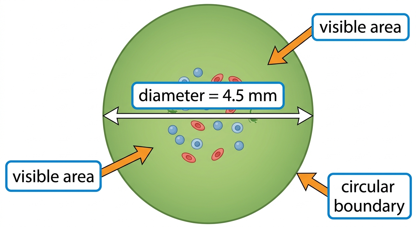
The diagram shows a circular field of view through a microscope. The diameter of this circle is mm.
a
Identify what "field of view" means when looking through a microscope. [1]
b
Describe how the diameter is used to measure the size of the visible area shown in the diagram. [1]
Solutions
10QuestionUsing a Microscope to View Prepared SlidesAssessment Practice
5 marks~8 minCriterion C
Three students each measured the length of the same onion cell four times. The accepted length is 125 μm.
| Student A | 125 μm | 127 μm | 124 μm | 126 μm |
|---|---|---|---|---|
| Student B | 120 μm | 130 μm | 122 μm | 128 μm |
| Student C | 115 μm | 115 μm | 116 μm | 114 μm |
a
Calculate the mean measurement for Student A. [1]
b
The range for Student C is 2 μm and the range for Student B is 10 μm. Describe what the range tells you about Student C's measurements compared to Student B's. [2]
c
Student A's mean is 125.5 μm and Student B's mean is 125.0 μm. Student C's mean is 115.0 μm. The accepted length is 125 μm. Compare Student B's measurements with Student C's measurements. In your answer, say which student's measurements are more useful and why. [2]
Solutions
11QuestionMagnification and Field of ViewConcept Practice
2 marks~3 minCriterion A
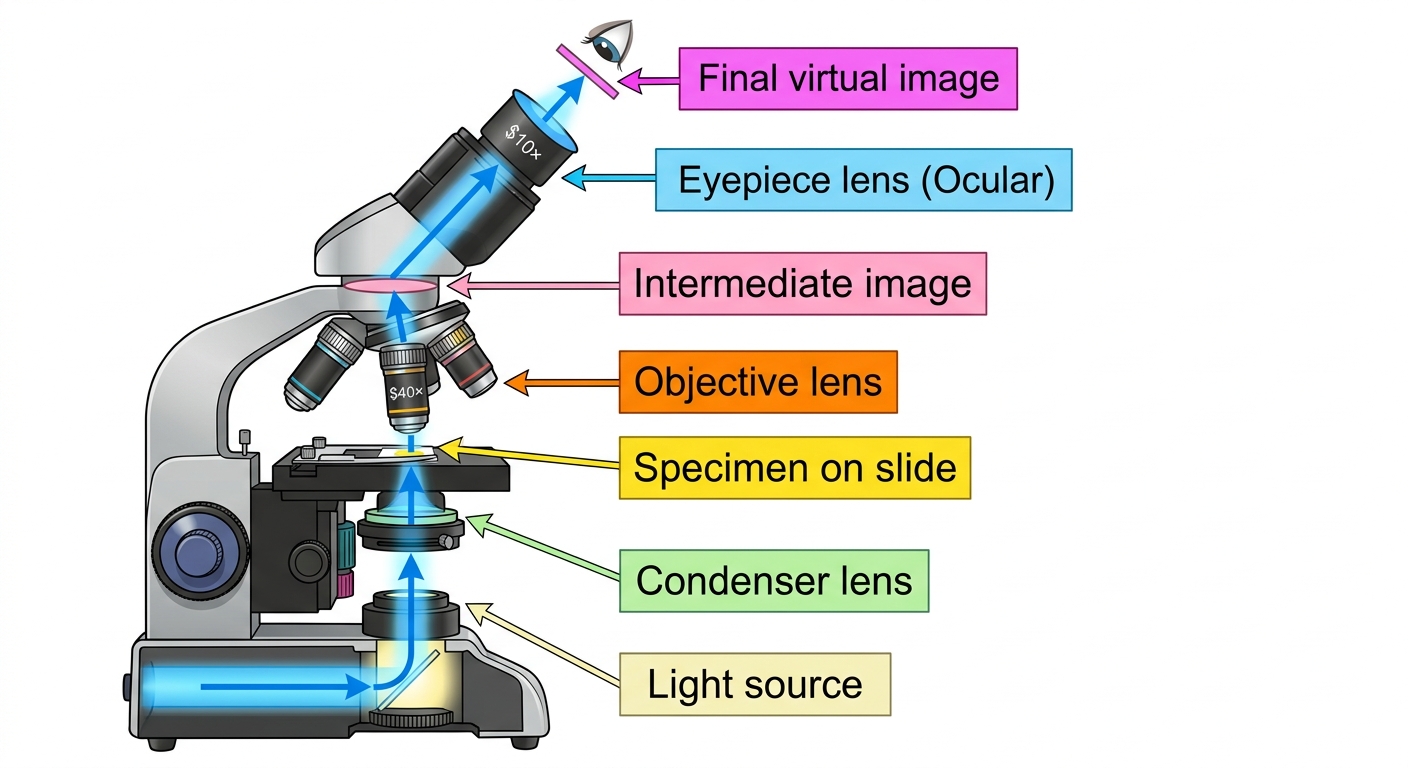
A microscope has an eyepiece lens with a magnification of and an objective lens with a magnification of .
Calculate the total magnification of the microscope when both lenses are used together. [2]
Calculate the total magnification of the microscope when both lenses are used together. [2]
Solutions
12QuestionSafety and Handling of MicroscopesAssessment Practice
5 marks~8 minCriterion B
A class recorded five microscope accidents.
Accident 1 — Cause: carried with one hand → Consequence: microscope fell
Accident 2 — Cause: used high power without checking → Consequence: lens scratched
Accident 3 — Cause: touched lens with fingers → Consequence: smudge on lens
Accident 4 — Cause: did not secure slide → Consequence: slide broke
Accident 5 — Cause: pulled plug by the cord → Consequence: wire damaged
Accident 6 — Cause: did not lower the stage before removing the slide
Accident 1 — Cause: carried with one hand → Consequence: microscope fell
Accident 2 — Cause: used high power without checking → Consequence: lens scratched
Accident 3 — Cause: touched lens with fingers → Consequence: smudge on lens
Accident 4 — Cause: did not secure slide → Consequence: slide broke
Accident 5 — Cause: pulled plug by the cord → Consequence: wire damaged
Accident 6 — Cause: did not lower the stage before removing the slide
a
Identify one unsafe action from the five accidents above that caused damage to a slide. [1]
b
Describe what happens to the slide in Accident 6 when the stage is not lowered before the slide is removed. [2]
c
State the most likely consequence for Accident 6 and explain how the pattern from all five accidents supports your answer. [2]
Solutions
13QuestionMagnification and Field of ViewConcept Practice
2 marks~3 minCriterion A
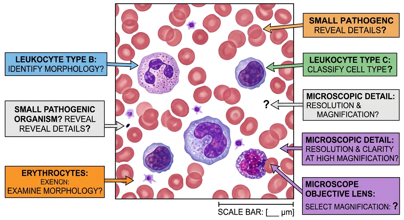
A doctor looks at a patient's blood sample under a microscope to check for signs of infection.
a
Identify one type of tiny living thing a doctor might find in an infected blood sample. [1]
b
Describe why using the correct magnification helps the doctor make the right decision about treatment. [1]
Solutions
14QuestionUsing a Microscope to View Prepared SlidesAssessment Practice
6 marks~9 minCriterion A
You used a microscope to look at a prepared slide of Elodea leaf cells. At 100× magnification, you could see 8 cells across the diameter of the field of view.
a
Identify how many times greater 400× magnification is than 100× magnification. [1]
b
Describe what happens to the field of view when magnification increases. [2]
c
State how many Elodea leaf cells would be visible across the diameter at 400× magnification, and explain why. [3]
Solutions
15QuestionMaking Temporary Slides Onion Cells etcConcept Practice
2 marks~3 minCriterion D
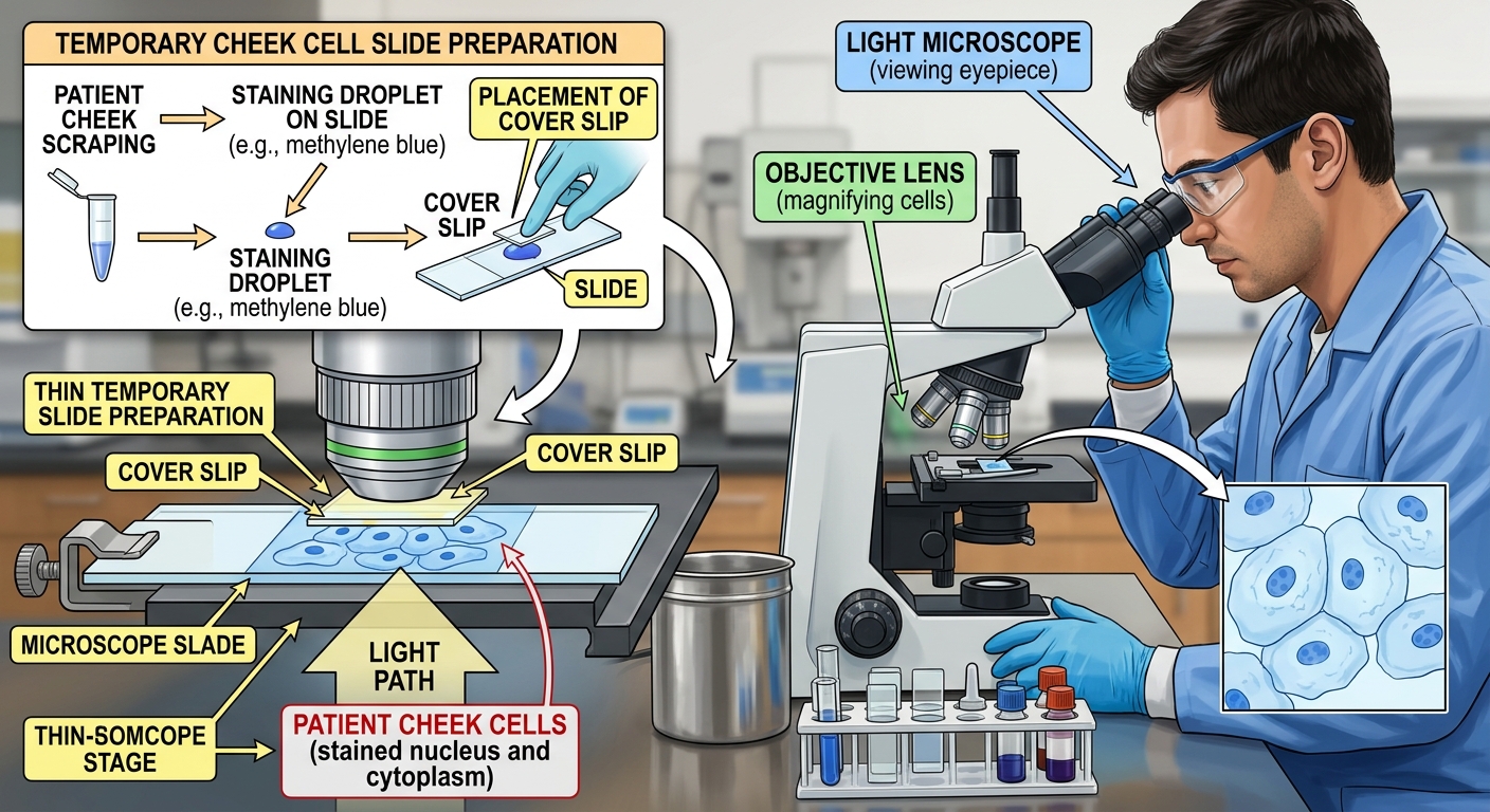
A doctor uses a microscope to look at cheek cells taken from a patient to check for signs of infection.
a
State one reason why the layer of cheek cells on the slide must be thin. [1]
b
Describe how a thin, clear slide helps the doctor care for the patient. [1]
Solutions
16QuestionUsing a Microscope to View Prepared SlidesAssessment Practice
6 marks~9 minCriterion A
A microscope has a eyepiece lens. Three objective lenses are available: , , and .
| Objective lens | |||
|---|---|---|---|
| Field of view diameter (mm) | 4.5 | 1.8 | 0.45 |
a
Identify the total magnification for each objective lens. Use the formula:
[3]
[3]
b
Describe what happens to the field of view diameter as the total magnification increases. [2]
c
A student switches from the objective to the objective. Describe how the view through the eyepiece changes, giving one reason why. [1]
Solutions
17QuestionSafety and Handling of MicroscopesConcept Practice
3 marks~5 minCriterion A
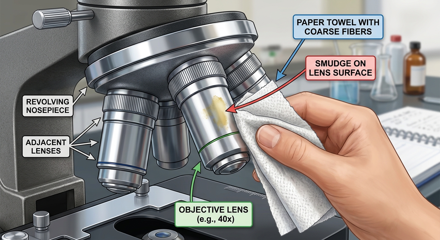
A student uses a paper towel to wipe a smudge off the objective lens of a microscope.
a
Name one reason why using a paper towel could harm the objective lens. [1]
b
Describe what damage this could cause to the microscope. [1]
c
State the correct materials a student should use to clean the objective lens safely. [1]
Solutions
18QuestionParts and Functions of a Light MicroscopeAssessment Practice
6 marks~9 minCriterion B
A student looks at cells using a light microscope. The table shows the objective lens and how small a detail the lens can show (called resolution).
| Objective lens | 4x | 10x | 40x | 100x |
|---|---|---|---|---|
| Resolution (µm) | 5.0 | 2.0 | 0.5 | 0.2 |
a
Name the pattern between the objective lens number and the resolution value shown in the table. [1]
b
Describe what resolution value you would expect for a 20x objective lens. Use the table to explain your answer. [2]
c
A plant cell is about 50 µm wide and a bacterium is about 1 µm wide. Describe why a scientist needs to use the 100x objective lens to see a bacterium, but only needs the 4x objective lens to see a plant cell. [3]
Solutions
19QuestionMaking Temporary Slides Onion Cells etcConcept Practice
4 marks~6 minCriterion A
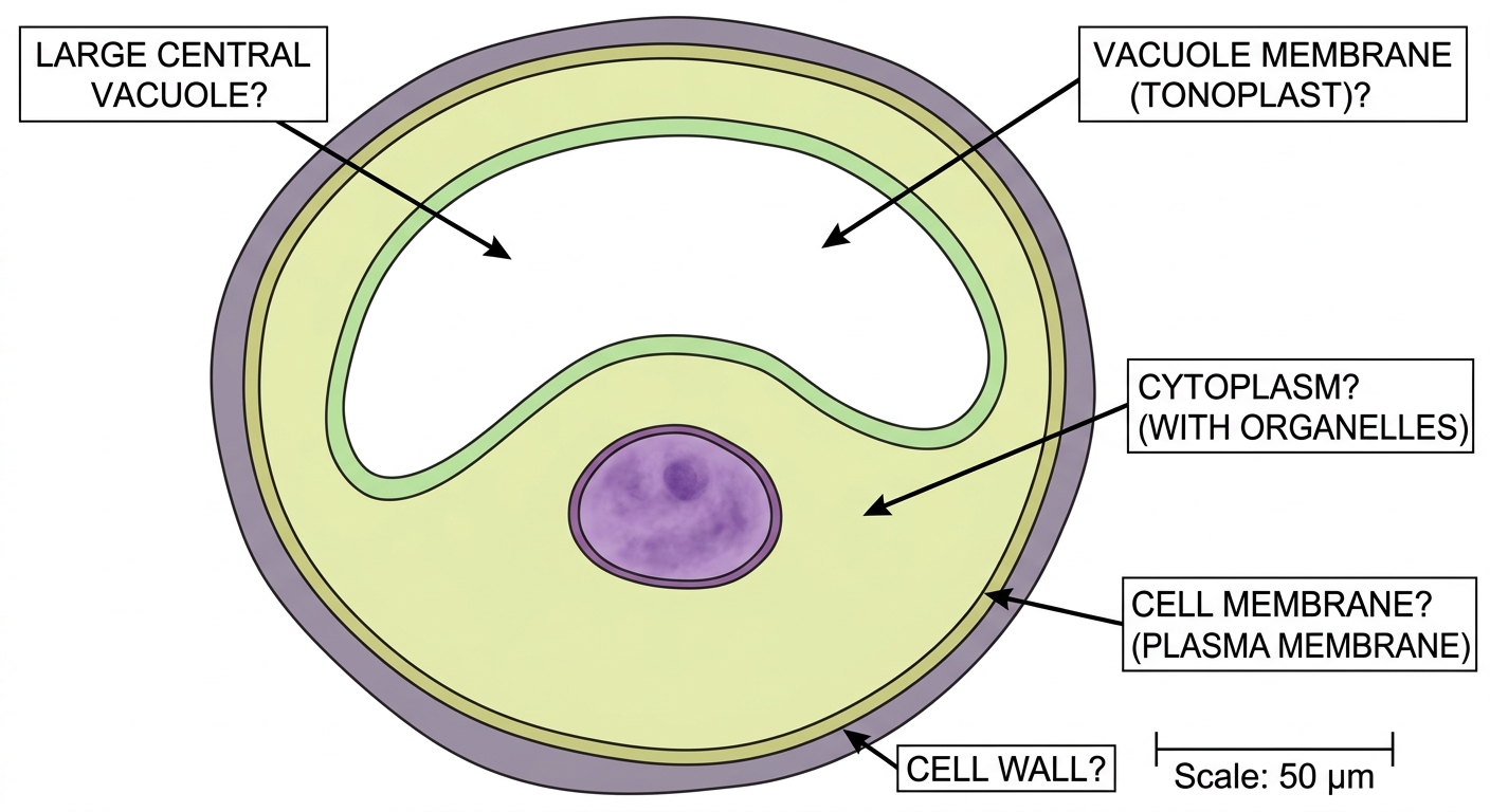
A student has drawn an onion cell diagram showing the cell wall and nucleus. The diagram has no labels yet.
a
Name one other part visible in an onion cell diagram. [1]
b
Describe two rules a student must follow when drawing a biological diagram. [2]
c
The student draws the cell membrane as a wavy, shaded line just inside the cell wall. Describe one way this is correct and one way this needs to be improved. [1]
Solutions
20QuestionSafety and Handling of MicroscopesAssessment Practice
6 marks~9 minCriterion C
Three students looked at the same onion skin slide under a microscope on high power. They each counted the cells they could see.
Student A: 12 cells — used the coarse focus knob on high power.
Student B: 8 cells — used the fine focus knob on high power.
Student C: 10 cells — cleaned the lens with a paper towel before looking.
Student A: 12 cells — used the coarse focus knob on high power.
Student B: 8 cells — used the fine focus knob on high power.
Student C: 10 cells — cleaned the lens with a paper towel before looking.
a
Identify which student used the correct focusing method on high power. [1]
b
Describe why one of the other students' results may not be reliable. [2]
c
Student B counted 8 cells. Students A and C counted different numbers. Describe two reasons why the three students got different counts from the same slide. [3]
Solutions
Showing 20 of 61 free questions