You're viewing free preview questions — 5 questions previewing here — subscribe to unlock 111 more, plus the Full Question Bank, Mock Papers & Worksheets.Upgrade
Skeletal System — Free MYP4 Biology Practice Questions

1QuestionProtection and support, homeostasisConcept Practice
2 marks~3 minCriterion D
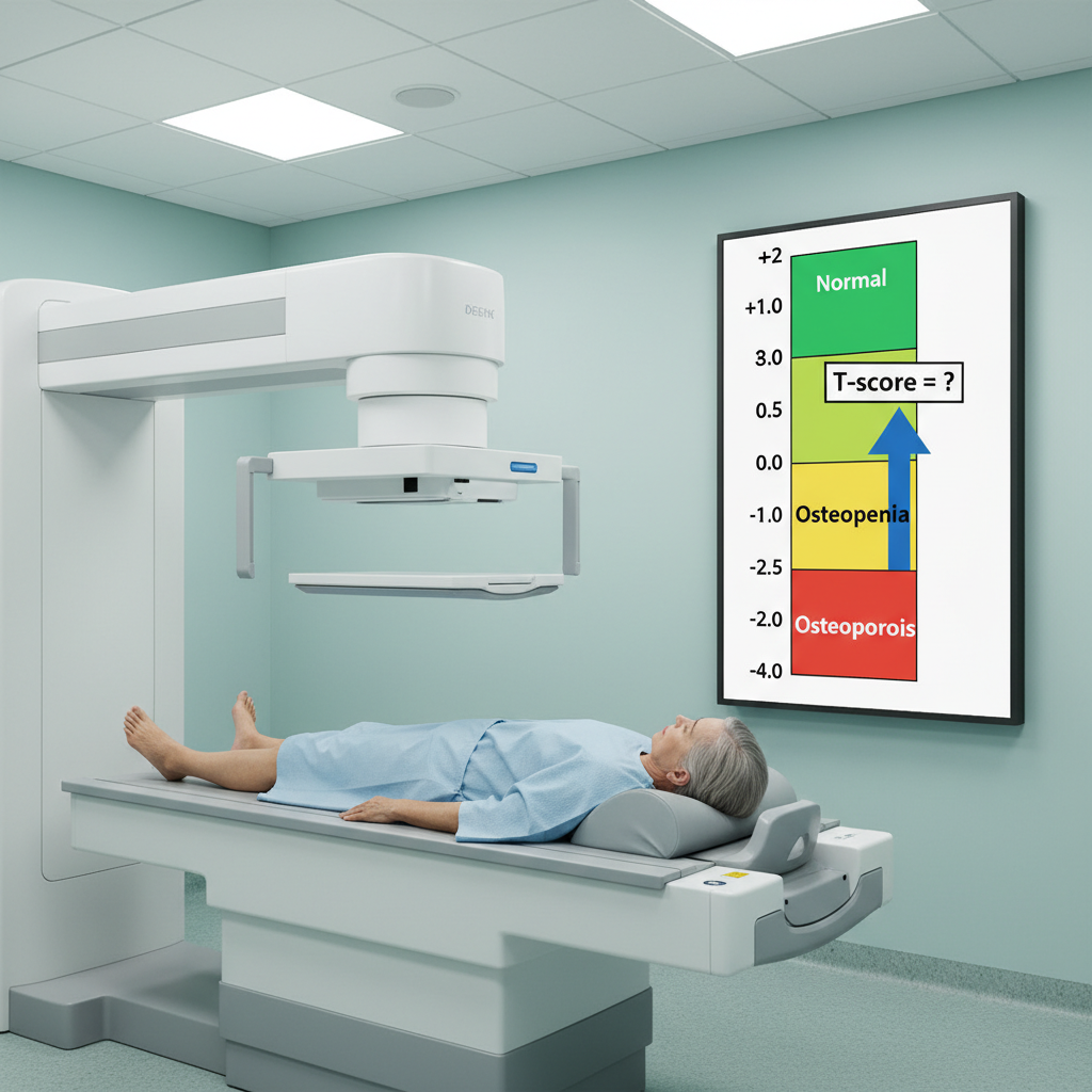
Bone density screening using dual-energy X-ray absorptiometry (DEXA) generates T-scores, which compare an individual's bone mineral density (BMD) to a healthy young-adult reference population. Clinicians use these scores to guide decisions about fracture prevention and treatment.
a
Outline one real-world medical application of bone density screening. [1]
b
Identify one limitation of relying on T-scores alone when assessing a patient's fracture risk. [1]

Solutions
2QuestionCommon problemsAssessment Practice
2 marks~3 minCriterion D
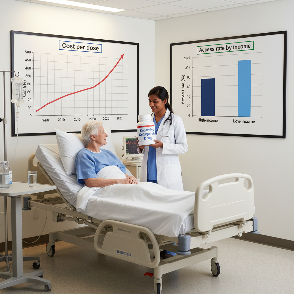
A pharmaceutical company develops a new drug that significantly reduces bone fractures in elderly patients with osteoporosis. The annual cost per patient is 12 000 dollars — unaffordable for many without comprehensive insurance.
Identify one ethical issue arising from the drug's high cost and its use in elderly patients, and explain how this issue affects healthcare equity. [2]
Identify one ethical issue arising from the drug's high cost and its use in elderly patients, and explain how this issue affects healthcare equity. [2]
Solutions
3QuestionMain partsConcept Practice
2 marks~3 minCriterion A
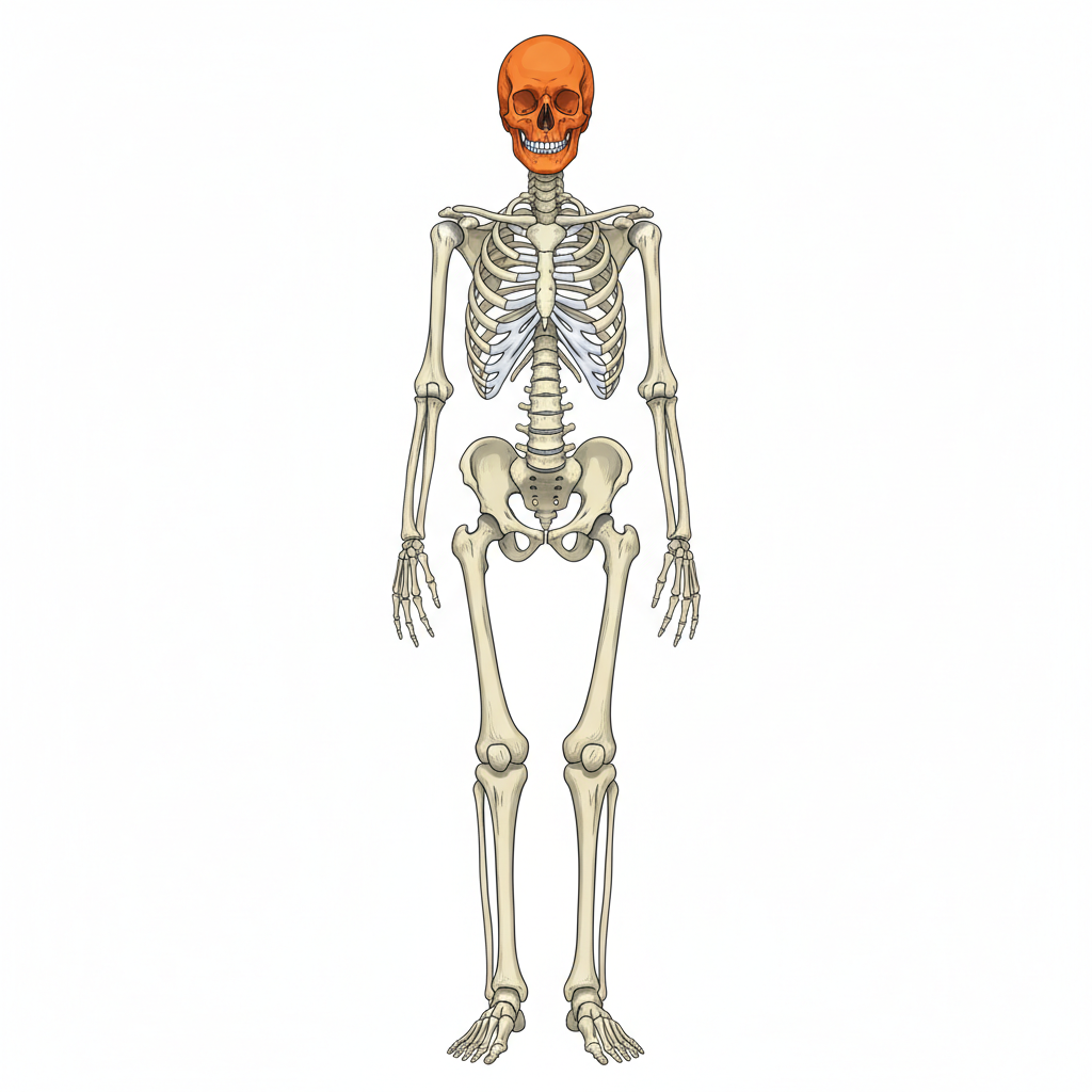
The diagram below shows the human skeleton with one region highlighted.
a
Identify the highlighted region of the skeleton. [1]
b
Explain one primary function of this region. [1]
Solutions
4QuestionWorking with musclesAssessment Practice
3 marks~5 minCriterion C
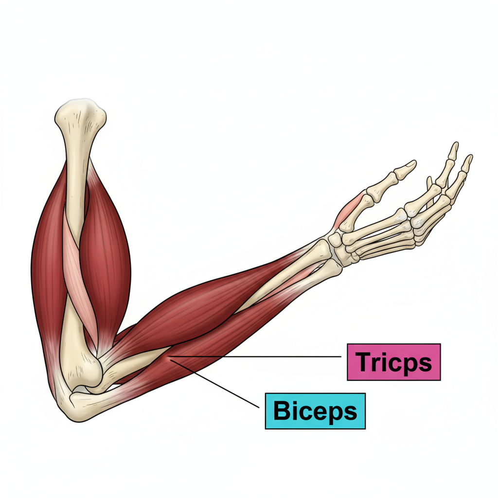
The diagram shows a human arm bent at 90° at the elbow. The biceps is shown contracted and bulging; the triceps is shown relaxed and elongated.
a
Identify which muscle is contracting and which is relaxing in the diagram. [1]
b
Explain how the biceps and triceps work together as an antagonistic pair to produce the bending movement shown. [1]
c
A person holds a heavy bag, keeping the arm bent at 90°. Evaluate whether the triceps is completely inactive in this position. [1]
Solutions
5QuestionMain partsAssessment Practice
6 marks~9 minCriterion A
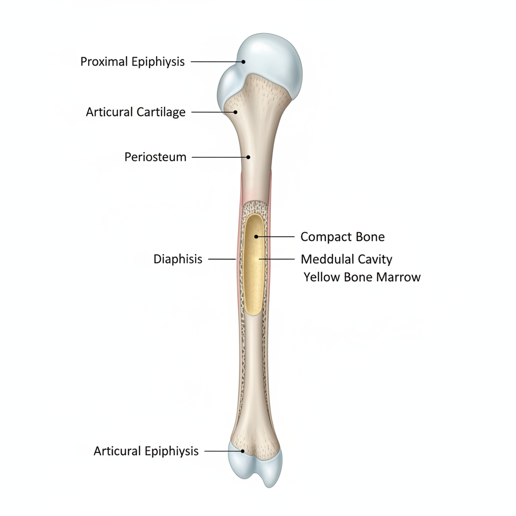
The diagram below shows a labelled femur with the following structures identified: proximal epiphysis, distal epiphysis, diaphysis (shaft), periosteum, compact bone, spongy bone, medullary cavity, and articular cartilage.
The table below shows average bone density measured at the femur neck in healthy adults:
The table below shows average bone density measured at the femur neck in healthy adults:
| Age group (years) | 20–29 | 30–39 | 40–49 | 50–59 | 60–69 | 70–79 |
|---|---|---|---|---|---|---|
| Bone density () | 0.95 | 0.93 | 0.90 | 0.85 | 0.78 | 0.70 |
a
Identify the bone layer visible in the diagram that primarily resists compressive forces during weight-bearing, and describe how its structure enables this function. [2]
b
Analyse the trend in bone density shown in the table and explain how the change observed after age 50 affects the femur's ability to support body weight. [2]
c
Justify why the medullary cavity is located in the diaphysis rather than the epiphyses, referring to the functional roles of both regions. [2]
Solutions
That's every free question here — 5 of 116.
+111 more questions, plus Mock Papers & Worksheets, are waiting in Premium.