Microscopes and Cell Observation — Free MYP2 Sciences Practice Questions

1QuestionParts and Functions of a Light MicroscopeConcept Practice
2 marks~3 minCriterion A
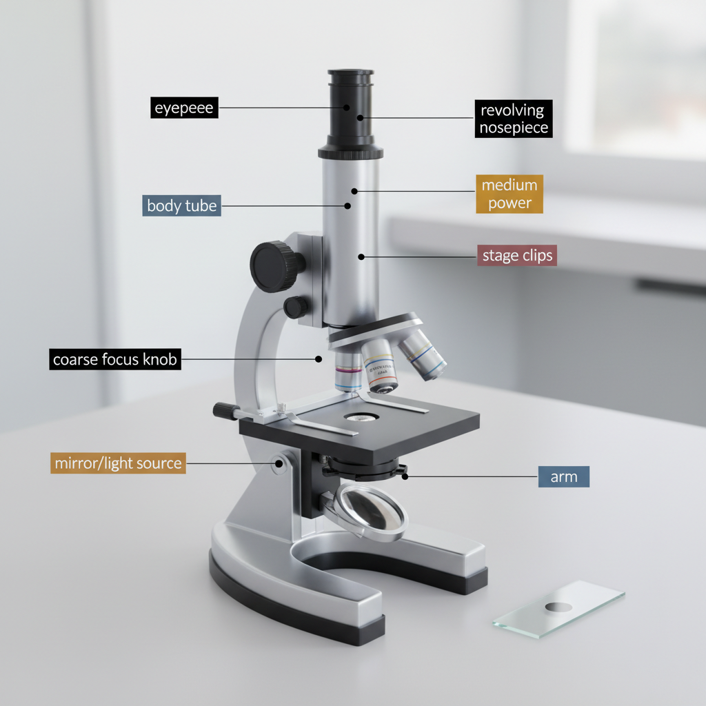
A student places a prepared slide on the stage of a light microscope and selects the lowest-power objective lens.
Describe the steps the student should follow, using the coarse focus knob and then the fine focus knob, to bring the specimen into clear focus. [2]
Describe the steps the student should follow, using the coarse focus knob and then the fine focus knob, to bring the specimen into clear focus. [2]

Solutions
2QuestionMagnification and Field of ViewAssessment Practice
2 marks~3 minCriterion A
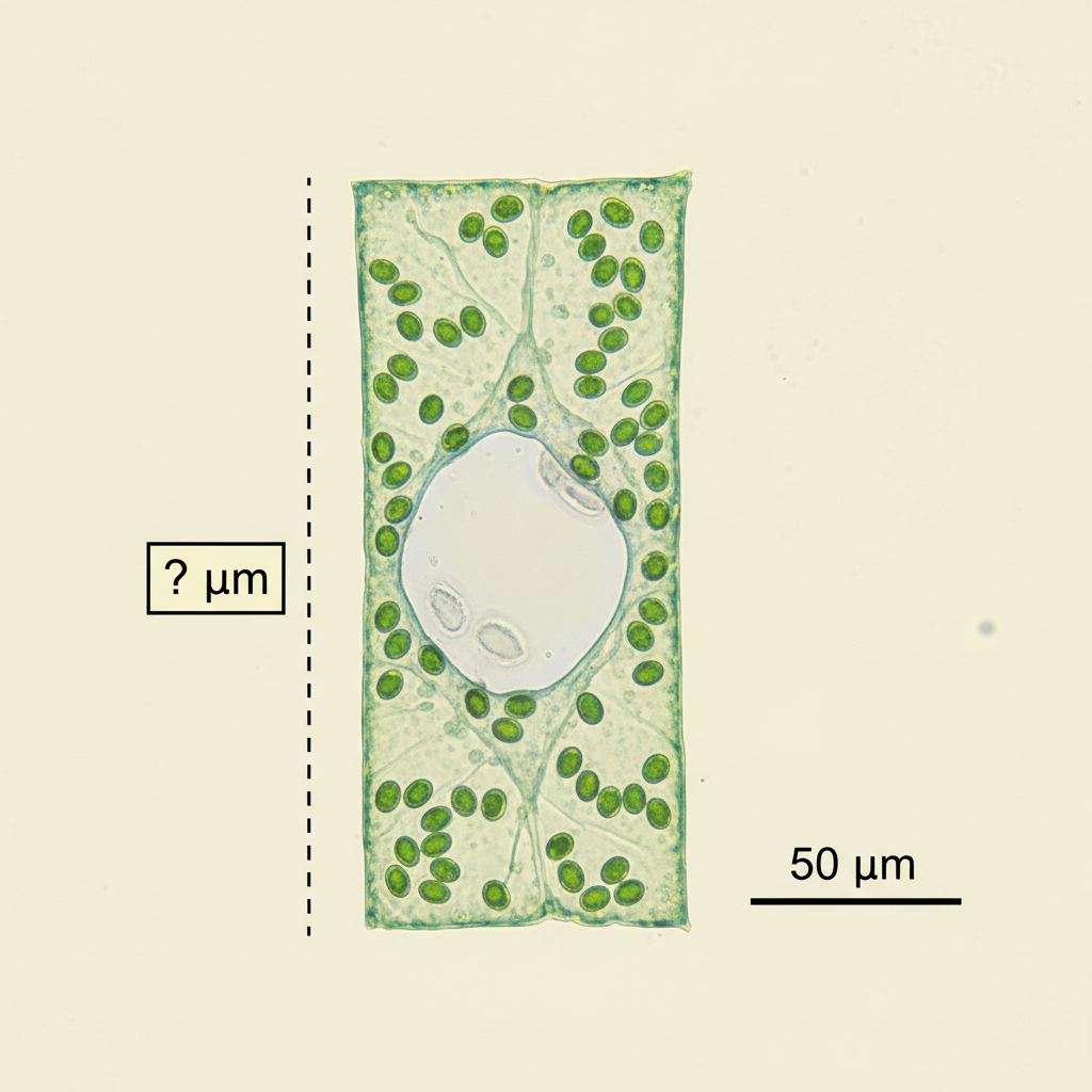
A micrograph of a plant cell includes a scale bar: the bar measures 1 cm on the image and represents an actual length of 50 μm (micrometres).
The cell measures 3 cm on the image.
Calculate the actual length of the cell in μm. Show your working. [2]
The cell measures 3 cm on the image.
Calculate the actual length of the cell in μm. Show your working. [2]
Solutions
3QuestionUsing a Microscope to View Prepared SlidesConcept Practice
2 marks~3 minCriterion A
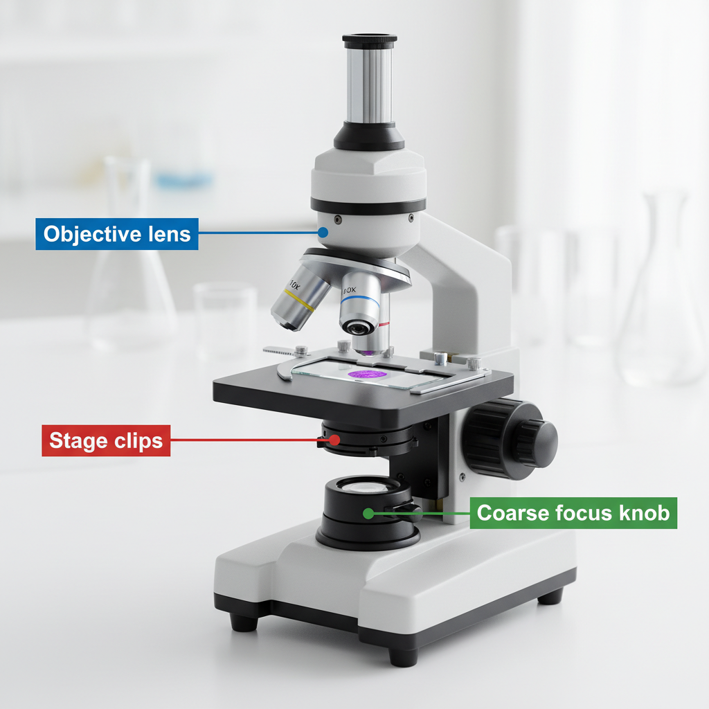
The diagram shows a microscope with three labeled parts: the objective lens, the stage clips, and the coarse focus knob.
a
Identify the function of the stage clips. [1]
b
Describe how the objective lens and the coarse focus knob work together to allow a student to view a specimen (a sample on a slide) clearly. [1]
Solutions
4QuestionMaking Temporary Slides Onion Cells etcAssessment Practice
2 marks~3 minCriterion A
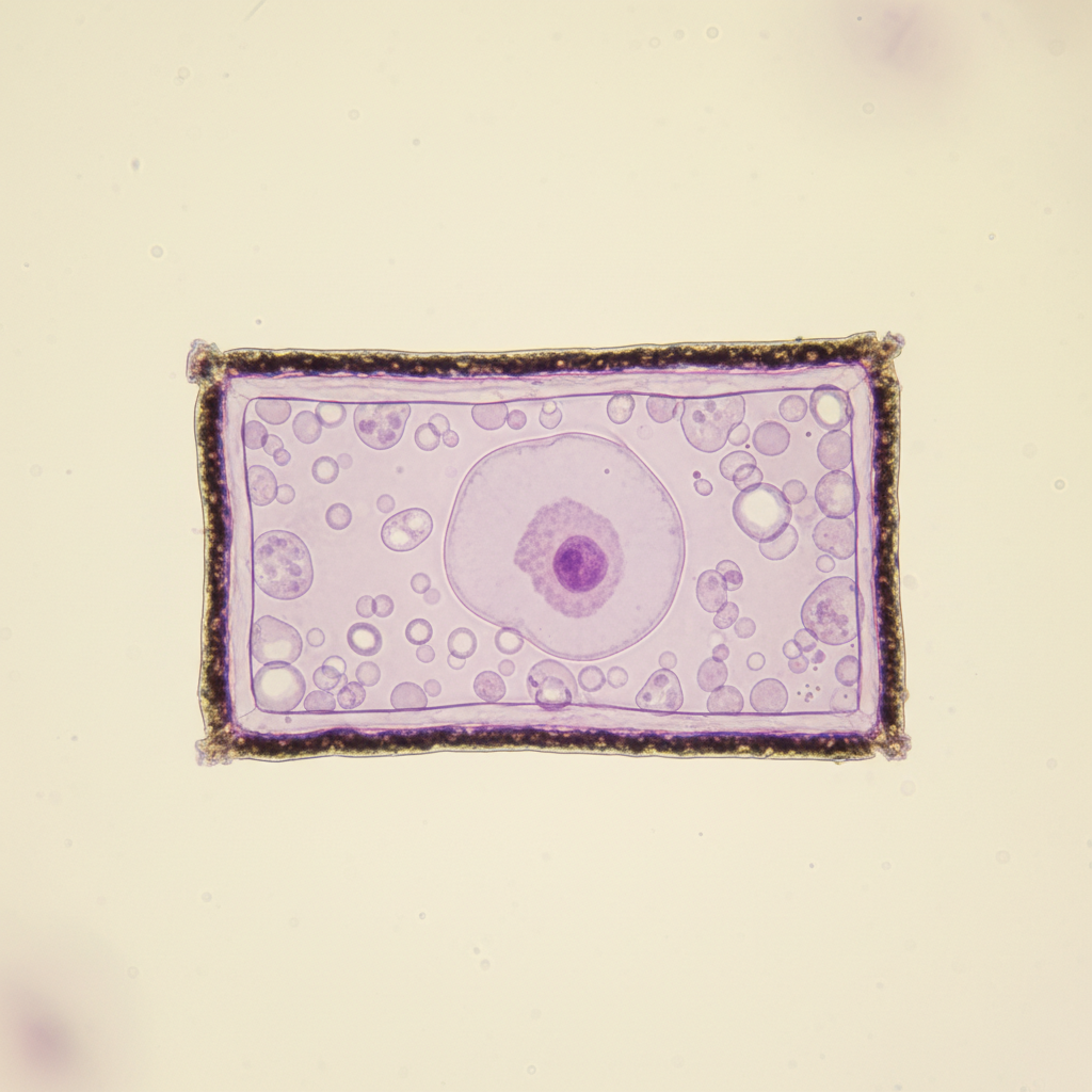
The diagram below shows a single onion cell stained with iodine solution. The cell wall (a thick, rigid layer made of cellulose) appears as a dark, thick line. The cell membrane (a thin, flexible layer just inside the wall) appears as a very faint line.
Explain why the cell wall is more visible than the cell membrane in this preparation. [2]
Explain why the cell wall is more visible than the cell membrane in this preparation. [2]
Solutions
5QuestionParts and Functions of a Light MicroscopeConcept Practice
2 marks~3 minCriterion A
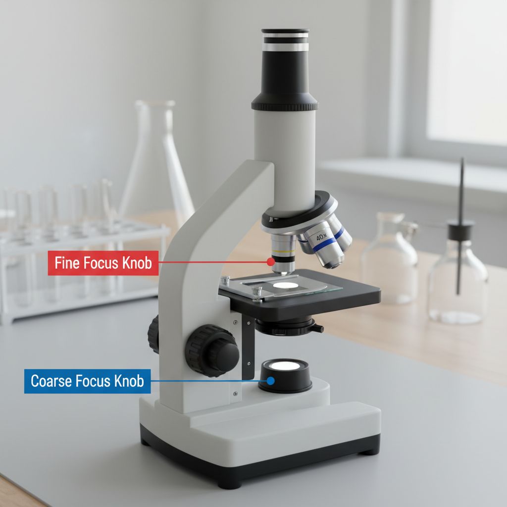
A biologist is examining living microorganisms (tiny organisms visible only under a microscope) in a drop of pond water. The organisms are actively swimming across the field of view.
a
Identify one reason why the biologist would use the fine focus knob rather than the coarse focus knob in this situation. [1]
b
Explain how using the fine focus knob helps the biologist keep the moving organisms in view. [1]
Solutions
6QuestionMaking Temporary Slides Onion Cells etcAssessment Practice
2 marks~3 minCriterion D
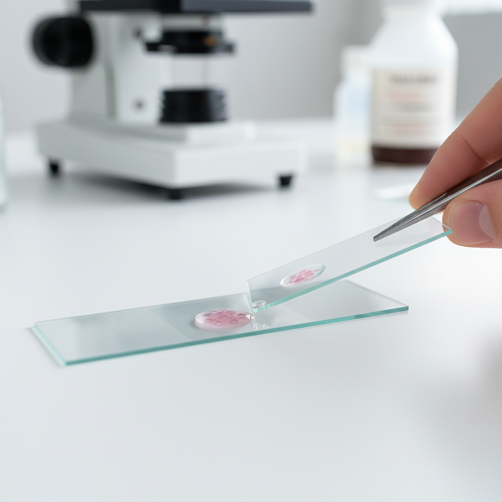
A medical technician prepares a temporary wet mount of a patient's cheek cells to check for infection.
Explain one reason why the technician should lower the coverslip slowly at an angle rather than dropping it flat onto the sample. [2]
Explain one reason why the technician should lower the coverslip slowly at an angle rather than dropping it flat onto the sample. [2]
Solutions
7QuestionSafety and Handling of MicroscopesConcept Practice
2 marks~3 minCriterion A
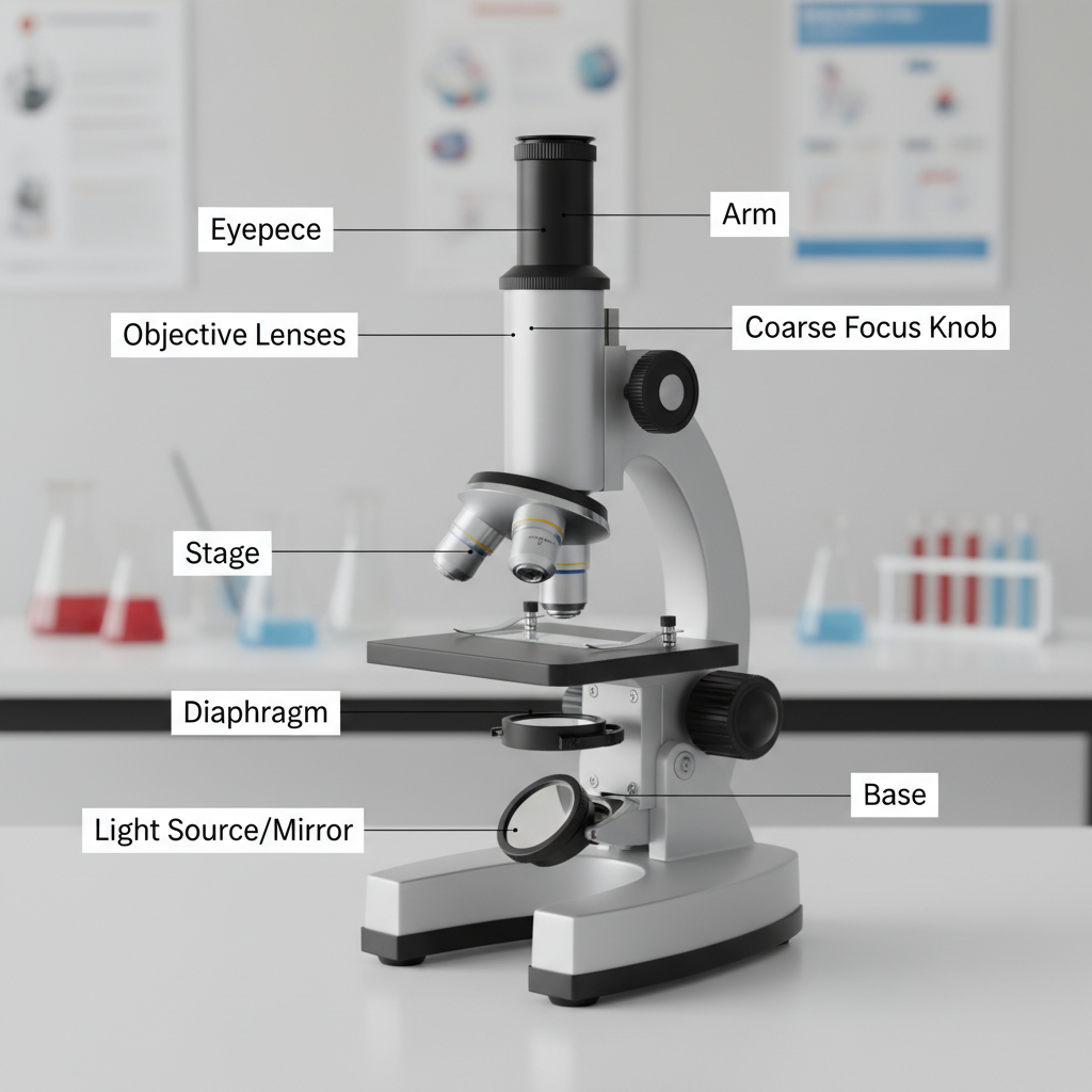
The diagram shows a labelled microscope.
a
Identify the part labelled 'coarse focus knob'. [1]
b
State one function of the coarse focus knob. [1]
Solutions
8QuestionMaking Temporary Slides Onion Cells etcAssessment Practice
3 marks~5 minCriterion A
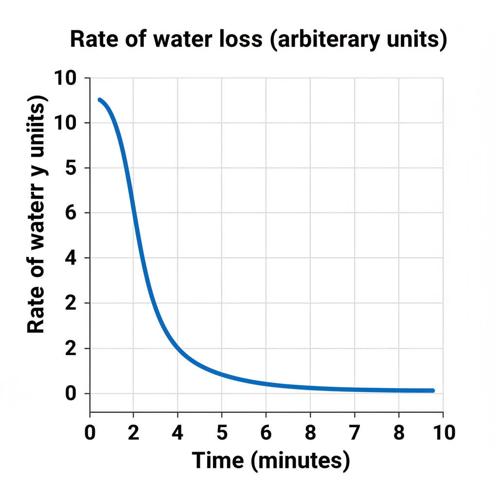
The graph below shows the rate of water loss from an onion cell placed in a concentrated salt solution (a solution with more dissolved salt than the cell contains).
a
Identify the trend shown in the graph. [1]
b
Explain why the rate of water loss is high at the start. [1]
c
Explain why the rate of water loss eventually reaches zero. [1]
Solutions
9QuestionParts and Functions of a Light MicroscopeConcept Practice
2 marks~3 minCriterion A
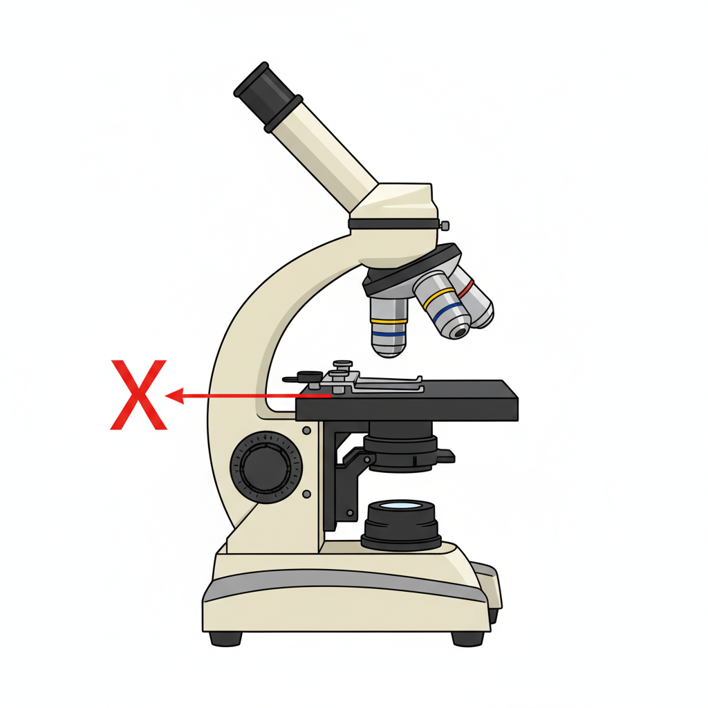
The diagram below shows a light microscope with part X labelled.
a
Identify part X. [1]
b
Describe the function of part X. [1]
Solutions
10QuestionUsing a Microscope to View Prepared SlidesAssessment Practice
3 marks~5 minCriterion B
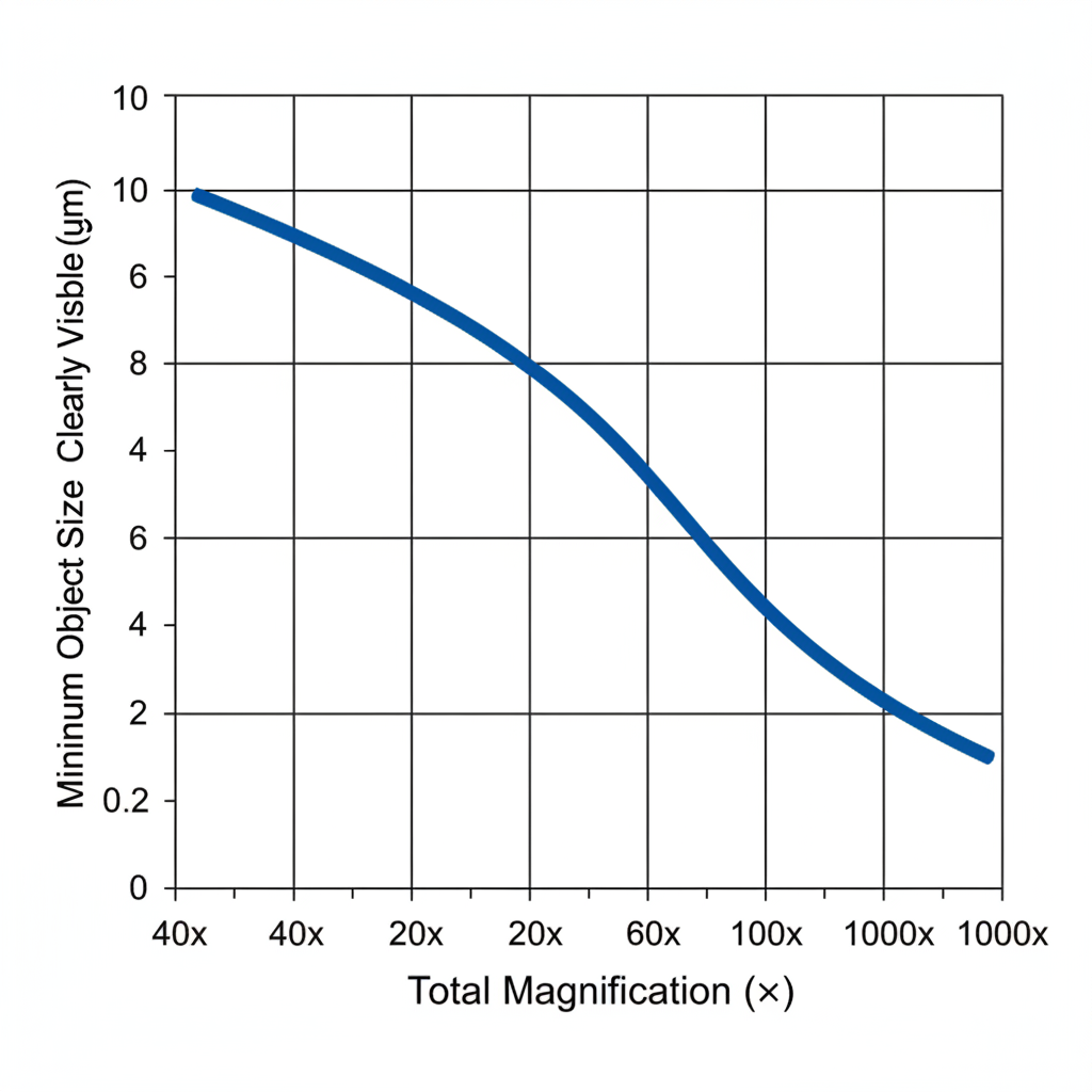
The graph below shows how total magnification affects the actual size of an object that can be clearly seen. The eyepiece lens is fixed at 10×.
a
Identify the trend shown in the graph. [1]
b
Explain why this trend occurs. [1]
c
A student switches from the 10× objective lens to the 40× objective lens. Describe how this change affects what the student can see, and suggest one advantage and one disadvantage of using the higher magnification. [1]
Solutions
11QuestionSafety and Handling of MicroscopesConcept Practice
2 marks~3 minCriterion A
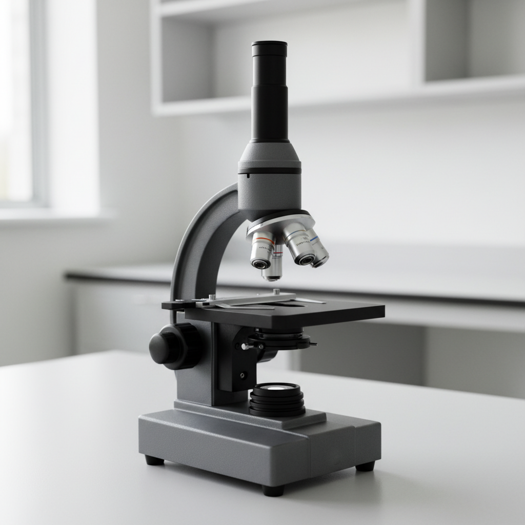
The diagram shows a microscope with its arm and base labelled.
Describe how to carry a microscope safely from a storage shelf to a lab bench. [2]
Describe how to carry a microscope safely from a storage shelf to a lab bench. [2]
Solutions
12QuestionSafety and Handling of MicroscopesAssessment Practice
3 marks~5 minCriterion C
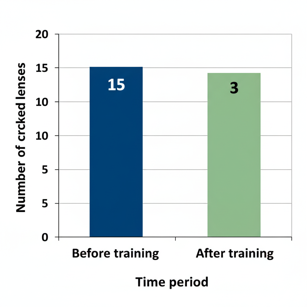
The bar graph below shows the number of cracked microscope lenses in a school laboratory during one term: 15 lenses cracked before safety training and 3 lenses cracked after safety training.
a
Identify the trend shown in the graph. [1]
b
Explain one reason why the number of cracked lenses decreased after safety training. [1]
c
A second school recorded 20 cracked lenses before training and 5 after. Compare the effectiveness of safety training in the two schools. [1]
Solutions
13QuestionUsing a Microscope to View Prepared SlidesConcept Practice
2 marks~3 minCriterion A
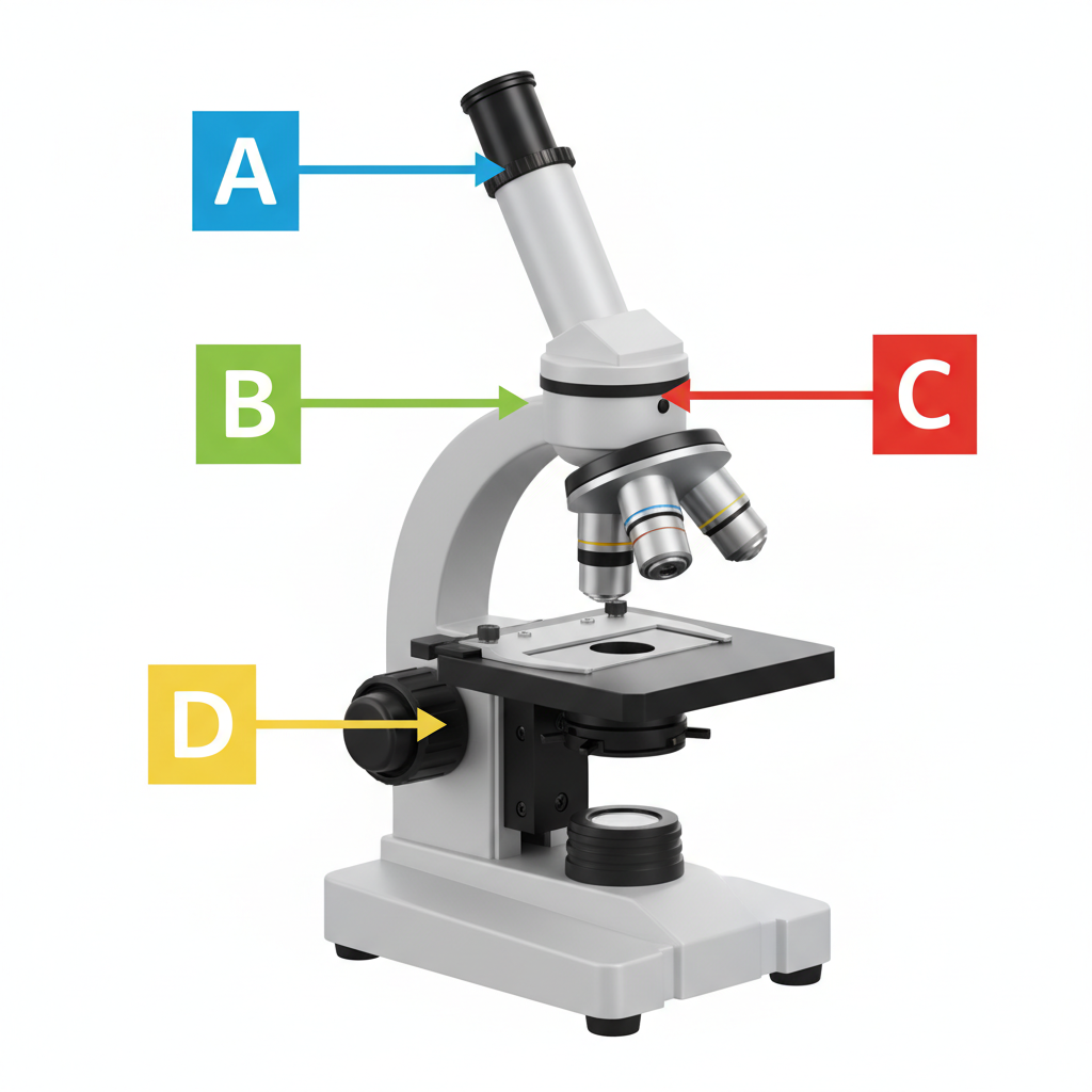
The diagram shows a microscope with four labeled parts: A (eyepiece), B (objective lens), C (stage), and D (coarse focus knob).
a
Identify the part used to bring the image into its first, rough focus. [1]
b
Explain why this part is used before any other focusing control when viewing a slide for the first time. [1]
Solutions
14QuestionUsing a Microscope to View Prepared SlidesAssessment Practice
4 marks~6 minCriterion A
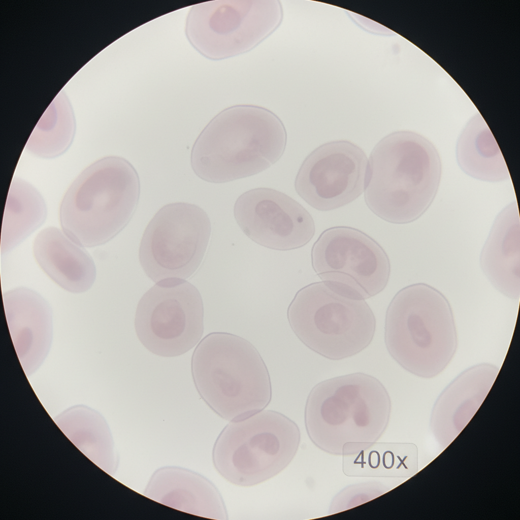
A student views a prepared slide of cheek cells under a light microscope at magnification. The image appears blurry, and the nucleus and cell membrane cannot be clearly seen.
a
Identify two limitations of using a light microscope that could cause this blurry image. [2]
b
Explain how using a stain (a dye that colours specific cell parts) could help overcome one of the limitations you identified in part (a). [2]
Solutions
15QuestionParts and Functions of a Light MicroscopeConcept Practice
2 marks~3 minCriterion A
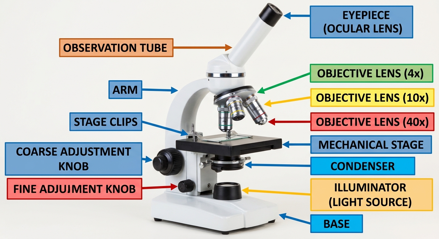
A light microscope has three objective lenses (the lenses closest to the specimen) labelled , , and .
a
Identify the objective lens that provides the highest magnification. [1]
b
Explain why that lens produces a higher magnification than the lens. [1]
Solutions
16QuestionParts and Functions of a Light MicroscopeAssessment Practice
4 marks~6 minCriterion A
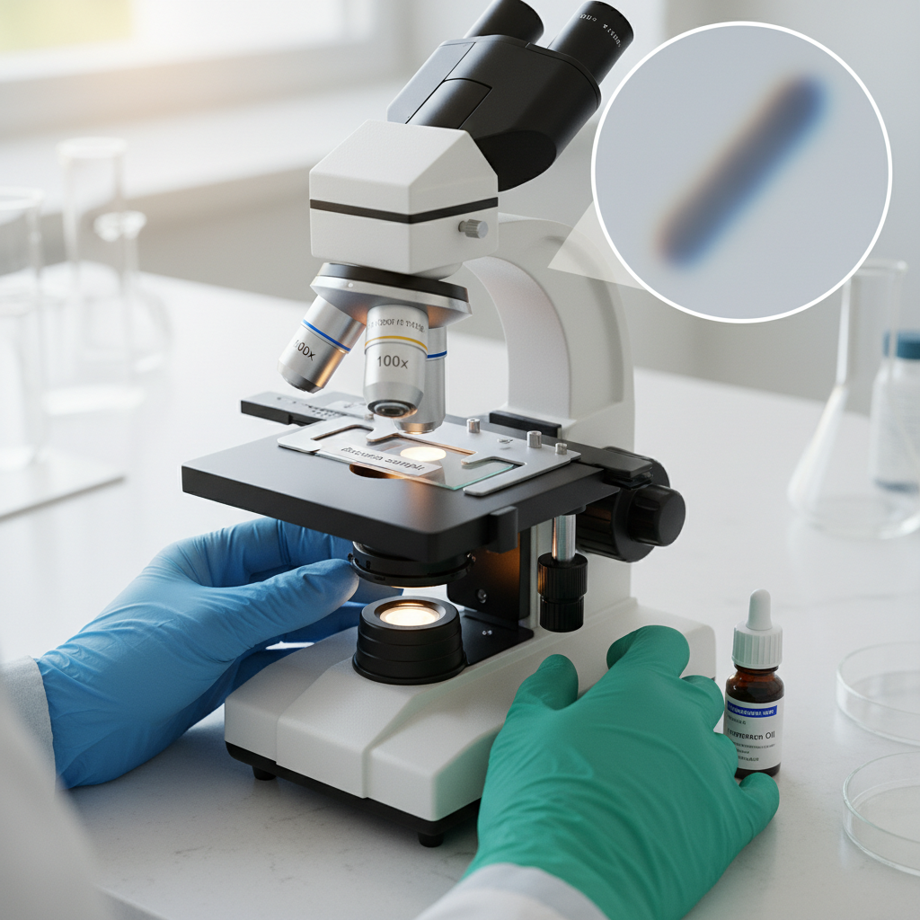
A student uses a light microscope to observe a bacterial cell. At a magnification of , the image appears blurry and the internal structures are not visible.
a
Identify the term that describes a microscope's ability to show two nearby structures as separate, and state the resolution limit of a light microscope. [1]
b
Explain why increasing magnification alone does not make the internal structures of the bacterial cell visible. [1]
c
Compare the use of an electron microscope with a light microscope for viewing bacterial internal structures, and identify one limitation of using an electron microscope. [2]
Solutions
17QuestionMagnification and Field of ViewConcept Practice
2 marks~3 minCriterion A
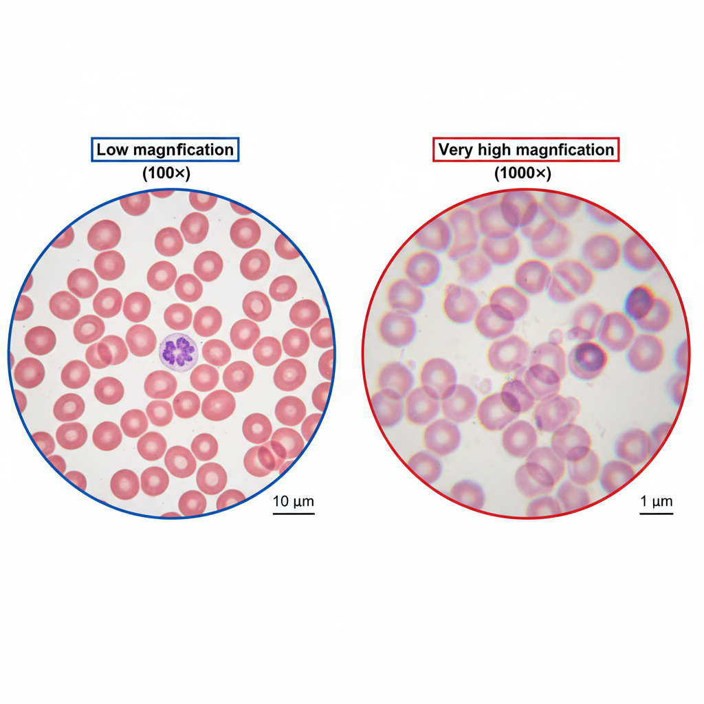
A doctor examines a patient's blood sample under a microscope to check for signs of disease.
a
Identify one reason why choosing the correct magnification is important for this examination. [1]
b
Explain one limitation of using excessively high magnification when viewing the blood sample. [1]
Solutions
18QuestionMaking Temporary Slides Onion Cells etcAssessment Practice
4 marks~6 minCriterion A
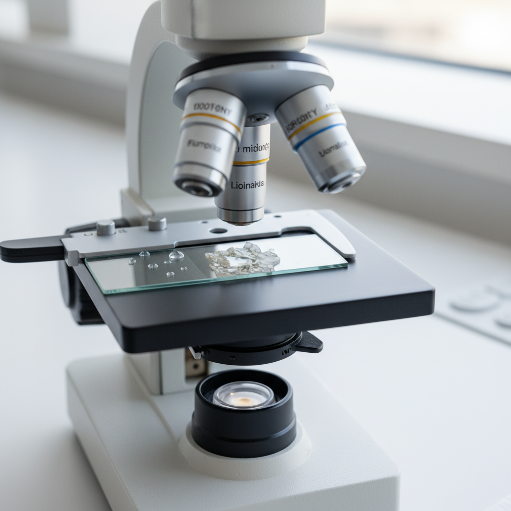
A student prepares a temporary slide of onion skin cells. When viewed under the microscope, air bubbles and overlapping cells are visible.
a
Identify one effect that air bubbles have on the appearance of cells. [1]
b
Explain how overlapping cells make it difficult to measure cell size accurately. [2]
c
Compare the reliability of observations made from this slide with those made from a correctly prepared slide. [1]
Solutions
19QuestionSafety and Handling of MicroscopesConcept Practice
2 marks~3 minCriterion D
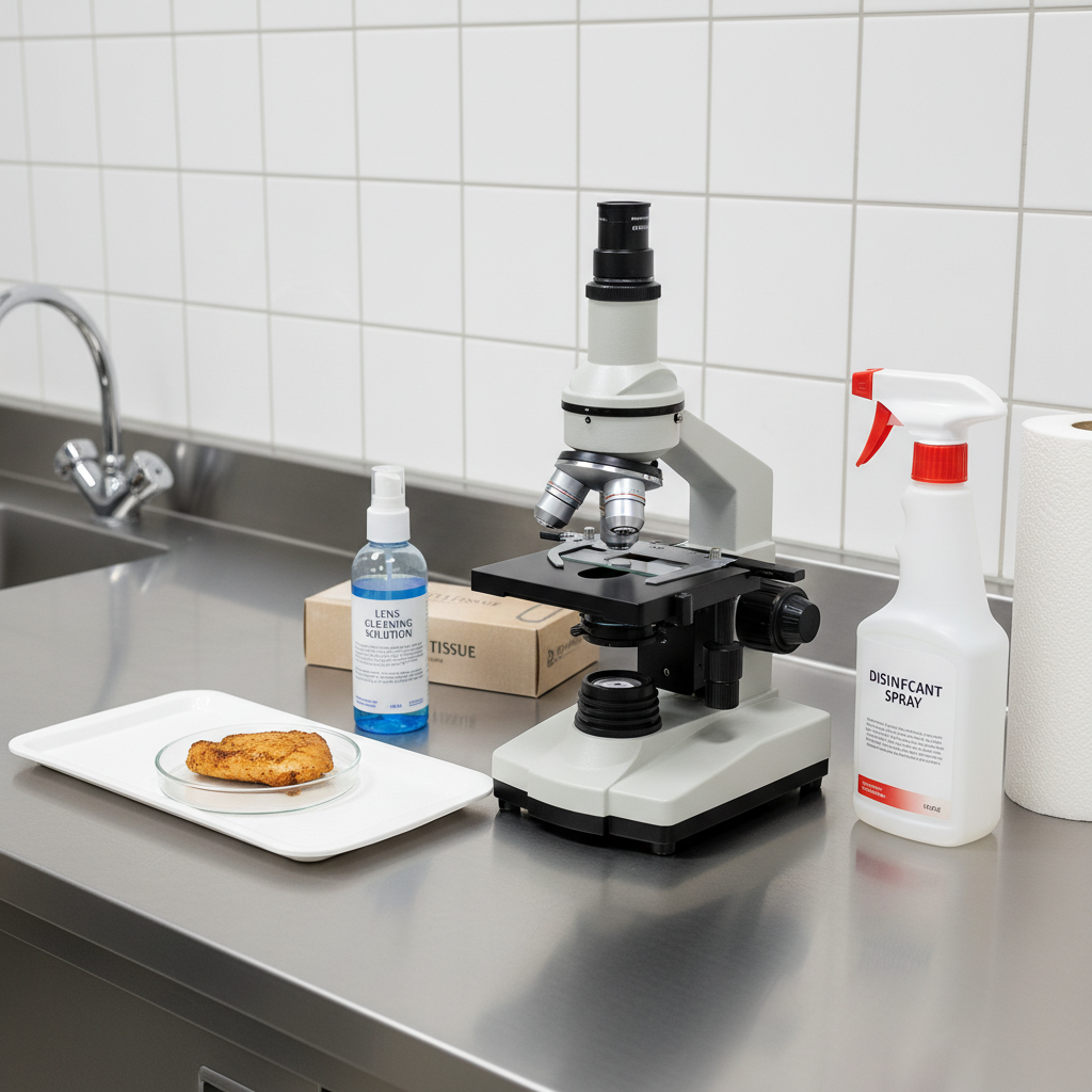
A hospital laboratory uses a microscope to check food samples for harmful bacteria (microorganisms that can cause illness).
a
Identify one cleaning or storage procedure that prevents contamination when using a microscope. [1]
b
Explain how this procedure helps prevent a health risk in the hospital. [1]
Solutions
20QuestionSafety and Handling of MicroscopesAssessment Practice
4 marks~6 minCriterion A
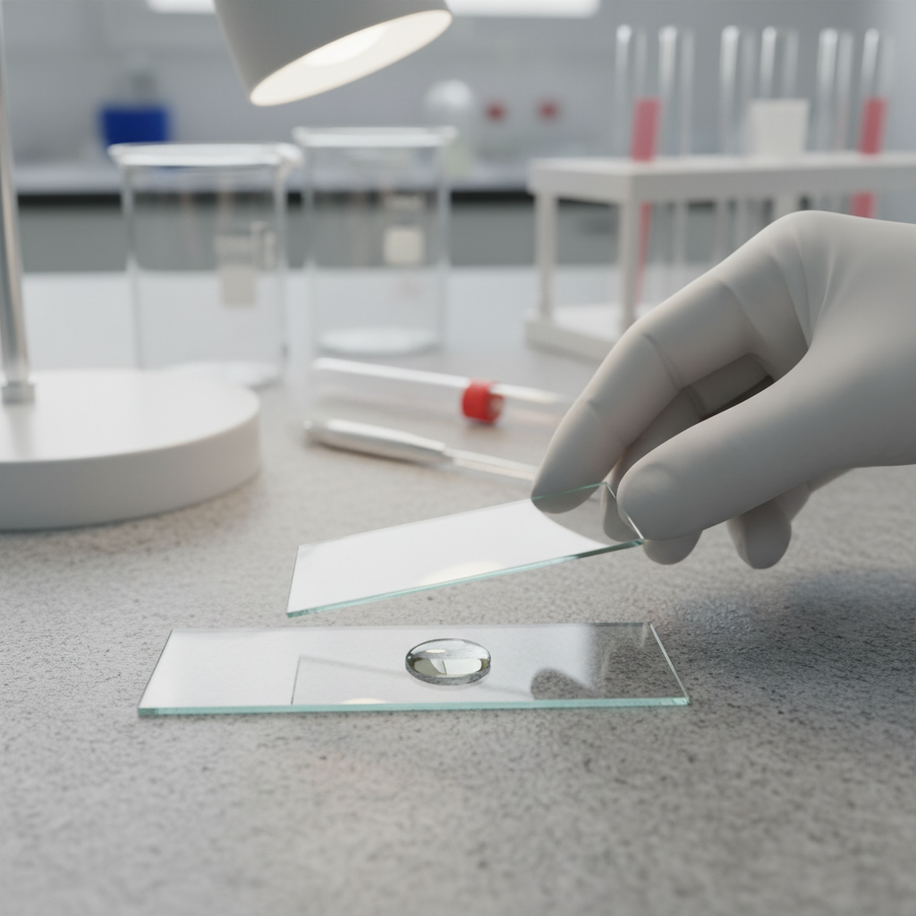
A student places a drop of water on a slide and lowers a cover slip flat onto it. The view shows a large, dark-edged circle that partly hides the specimen (the sample being observed).
a
Identify what the dark-edged circle is. [1]
b
Explain why the circle appears dark-edged. [1]
c
Describe the correct technique for placing a cover slip to prevent this problem. [2]
Solutions
Showing 20 of 81 free questions