Microscopes and Cell Observation — Free MYP3 Sciences Practice Questions

1QuestionUsing a Microscope to View Prepared SlidesConcept Practice
2 marks~3 minCriterion A
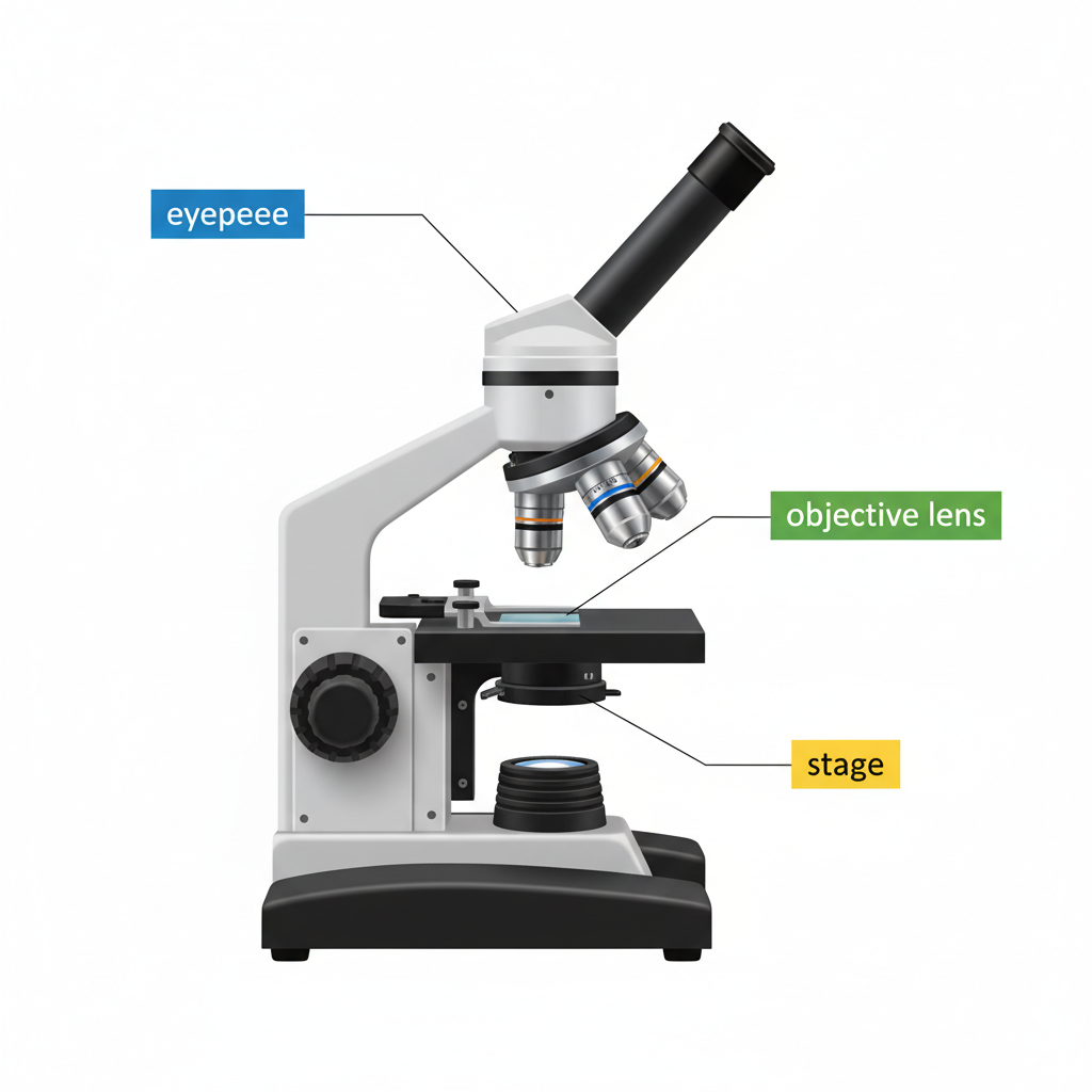
The diagram shows a microscope with three labeled components: the eyepiece, the objective lens, and the stage.
Describe the function of two of these three components. [2]
Describe the function of two of these three components. [2]

Solutions
2QuestionParts and Functions of a Light MicroscopeAssessment Practice
2 marks~3 minCriterion A
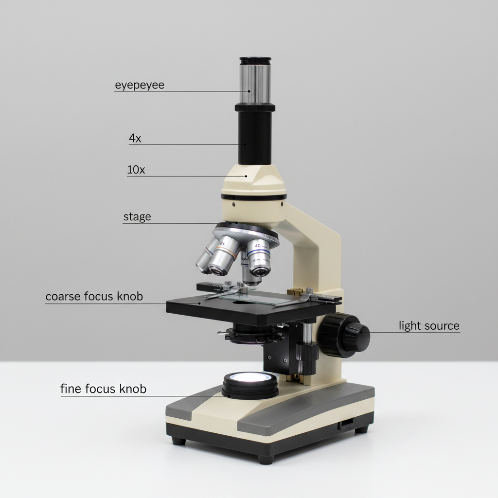
Describe the correct order of steps to bring a specimen into focus using a light microscope. Your answer must include all five terms: eyepiece, objective lens, stage, coarse focus knob, and fine focus knob. [2]
Solutions
3QuestionSafety and Handling of MicroscopesConcept Practice
2 marks~3 minCriterion A
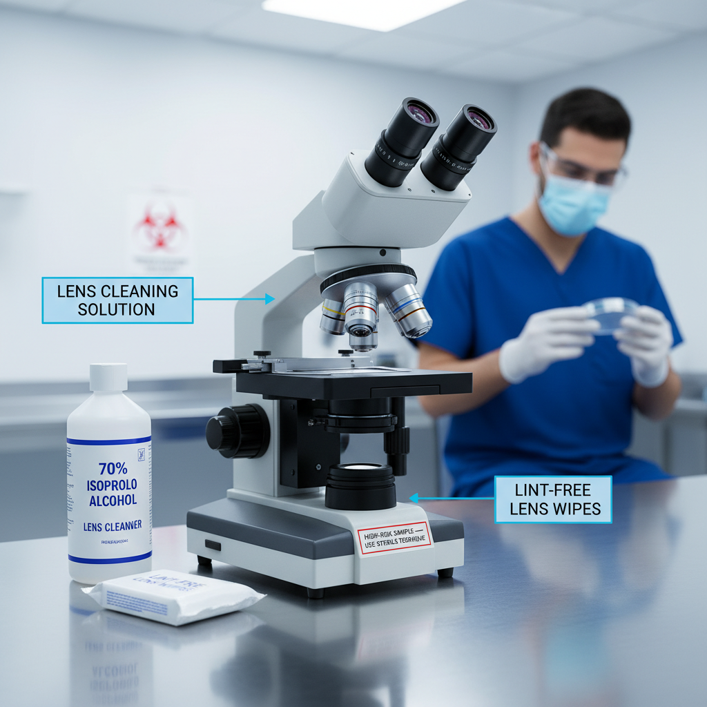
In a hospital laboratory, a technician examines blood samples from several patients using the same microscope during a single shift.
a
Describe one specific cleaning practice the technician should carry out between examining each patient's blood sample. [1]
b
Explain how this cleaning practice helps to prevent the spread of infection in the laboratory. [1]
Solutions
4QuestionMagnification and Field of ViewAssessment Practice
2 marks~3 minCriterion D
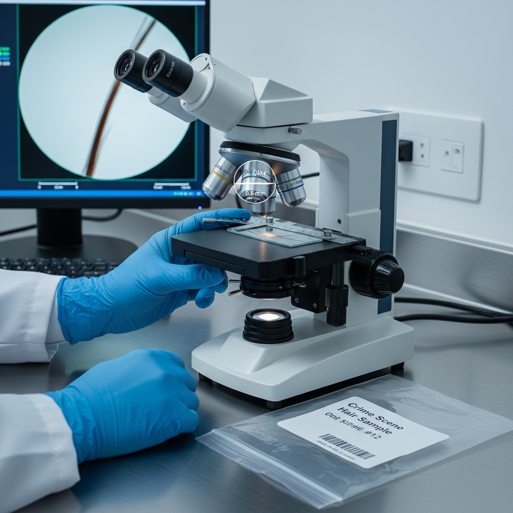
A forensic scientist uses a microscope to examine a hair sample found at a crime scene. The microscope has a magnification of ×400 and a field of view diameter of 0.5 mm.
Explain how knowing both the magnification and the field of view allows the forensic scientist to make useful measurements of the hair sample. [2]
Explain how knowing both the magnification and the field of view allows the forensic scientist to make useful measurements of the hair sample. [2]
Solutions
5QuestionUsing a Microscope to View Prepared SlidesConcept Practice
2 marks~3 minCriterion A
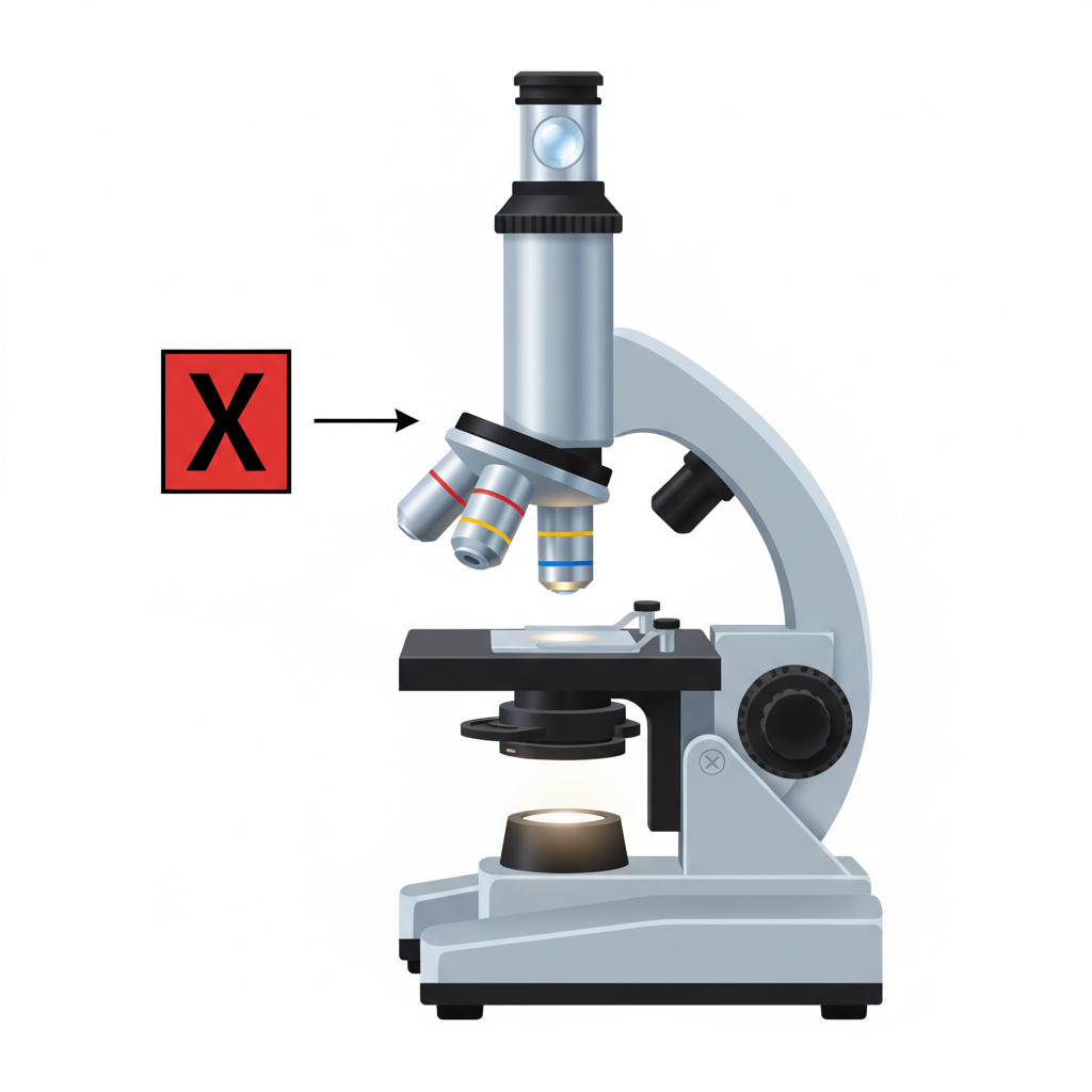
The diagram below shows a compound microscope with part X labelled.
a
Identify part X. [1]
b
Describe the function of part X in viewing a specimen. [1]
Solutions
6QuestionMagnification and Field of ViewAssessment Practice
3 marks~5 minCriterion C
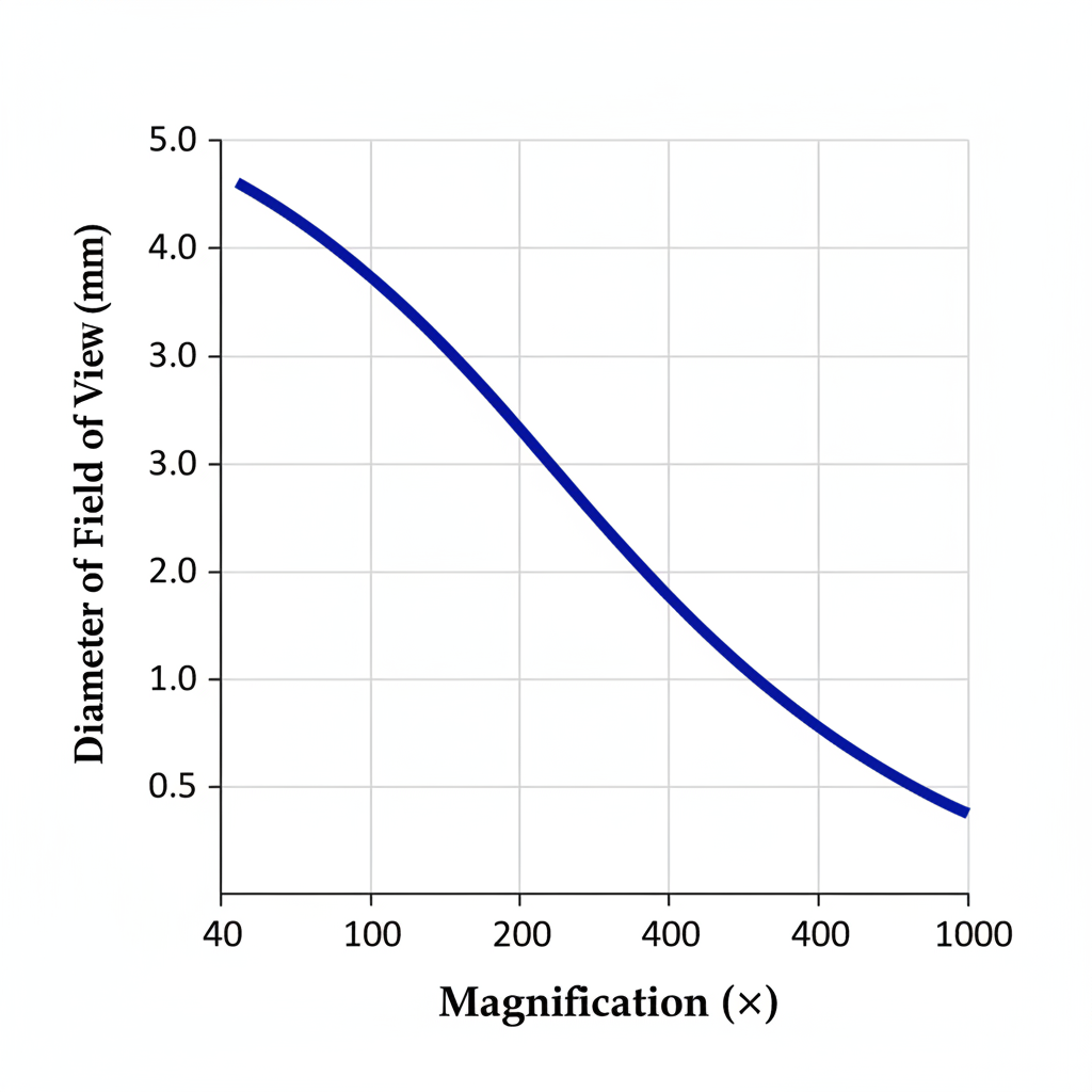
The graph below shows the relationship between the magnification of a microscope and the diameter of the field of view (in mm).
a
Describe the trend shown in the graph. [1]
b
Explain why the diameter of the field of view changes as magnification increases. [1]
c
A student claims that doubling the magnification will halve the diameter of the field of view. Using the graph, assess whether this claim is correct. [1]
Solutions
7QuestionParts and Functions of a Light MicroscopeConcept Practice
2 marks~3 minCriterion A
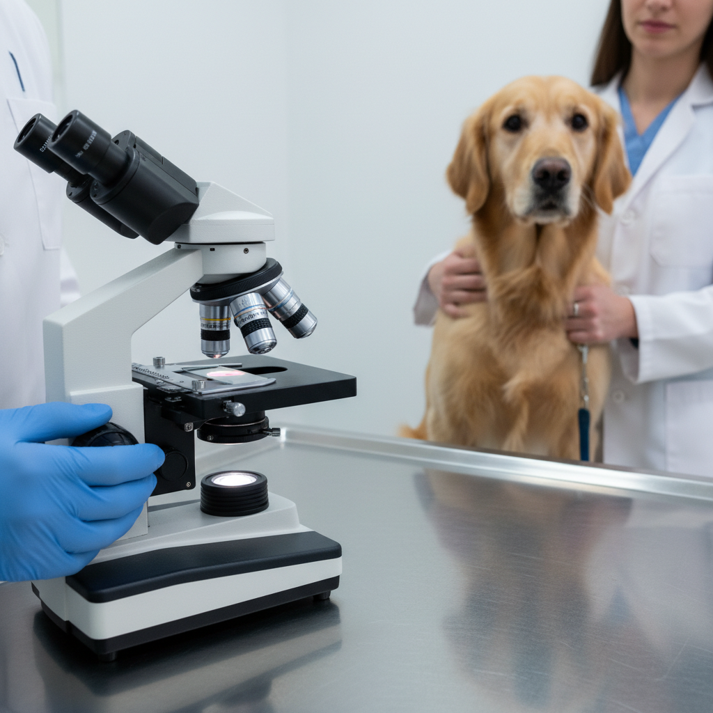
A veterinarian examines a dog's blood sample using a light microscope to check for parasites.
a
Describe one way a veterinarian uses a light microscope to examine a blood sample. [1]
b
Identify one limitation of using a light microscope for this type of examination. [1]
Solutions
8QuestionParts and Functions of a Light MicroscopeAssessment Practice
3 marks~5 minCriterion C
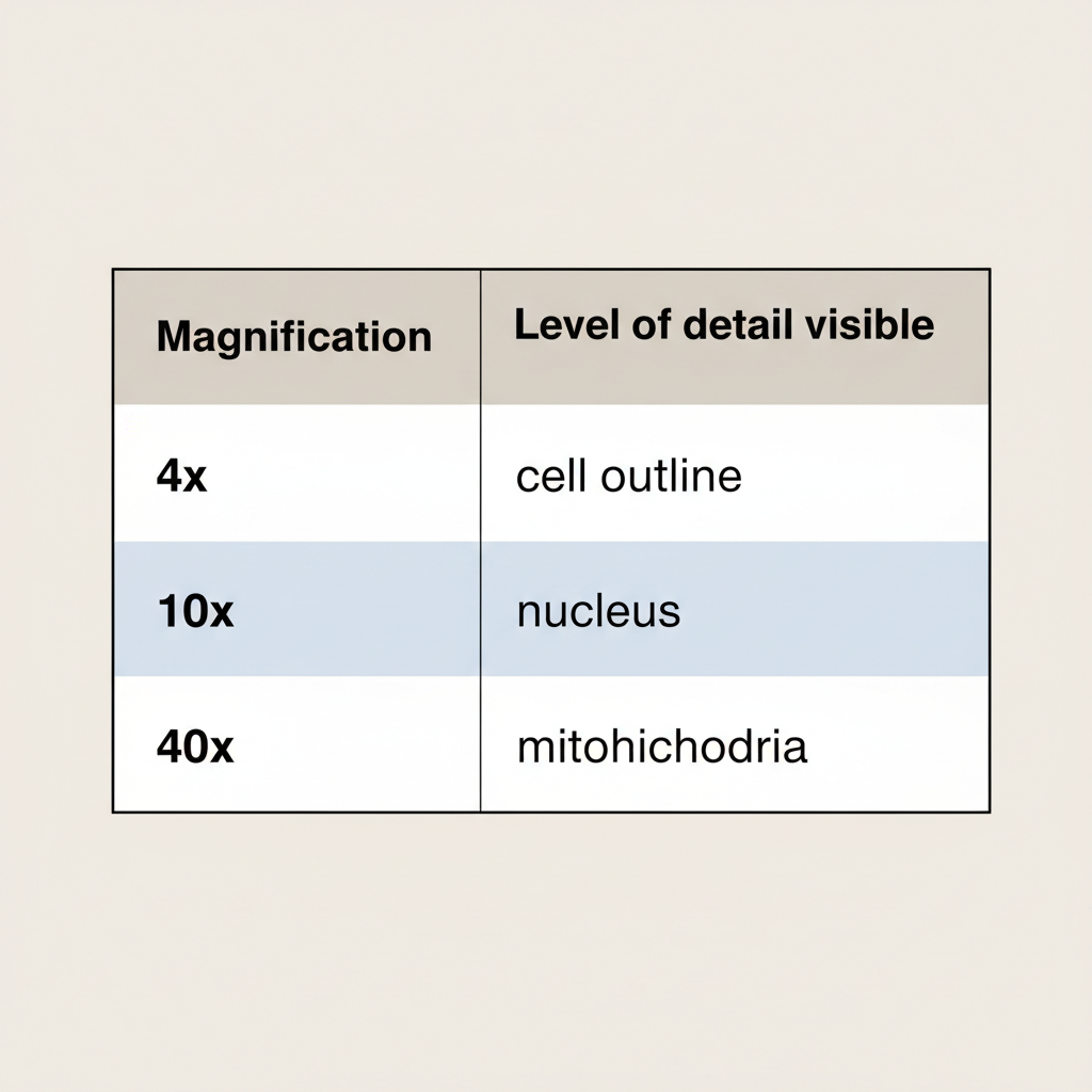
A student uses a light microscope at three different magnifications. The table below shows what becomes visible at each setting.
| Magnification () | 4 | 10 | 40 |
|---|---|---|---|
| Structure visible | cell outline | nucleus | mitochondria |
a
State the trend shown in the data. [1]
b
Explain why increasing magnification allows smaller cell structures to be seen. [1]
c
A classmate claims that magnification alone is enough to observe mitochondria clearly. Using the data and your knowledge, analyse whether this claim is fully correct. [1]
Solutions
9QuestionMaking Temporary Slides Onion Cells etcConcept Practice
2 marks~3 minCriterion A
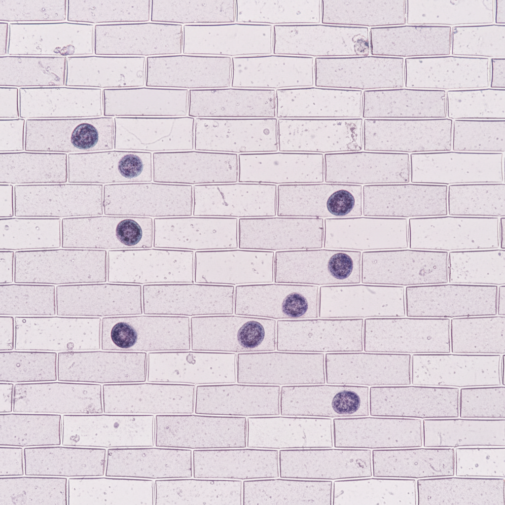
You have prepared a temporary slide of onion epidermal cells and are observing them under the microscope.
a
Describe the shape and arrangement of the onion cells you observe. [1]
b
Identify the three structures labelled in the cell diagram below — cell wall, nucleus, and cytoplasm — and describe the function of the nucleus. [1]
Solutions
10QuestionSafety and Handling of MicroscopesAssessment Practice
4 marks~6 minCriterion A
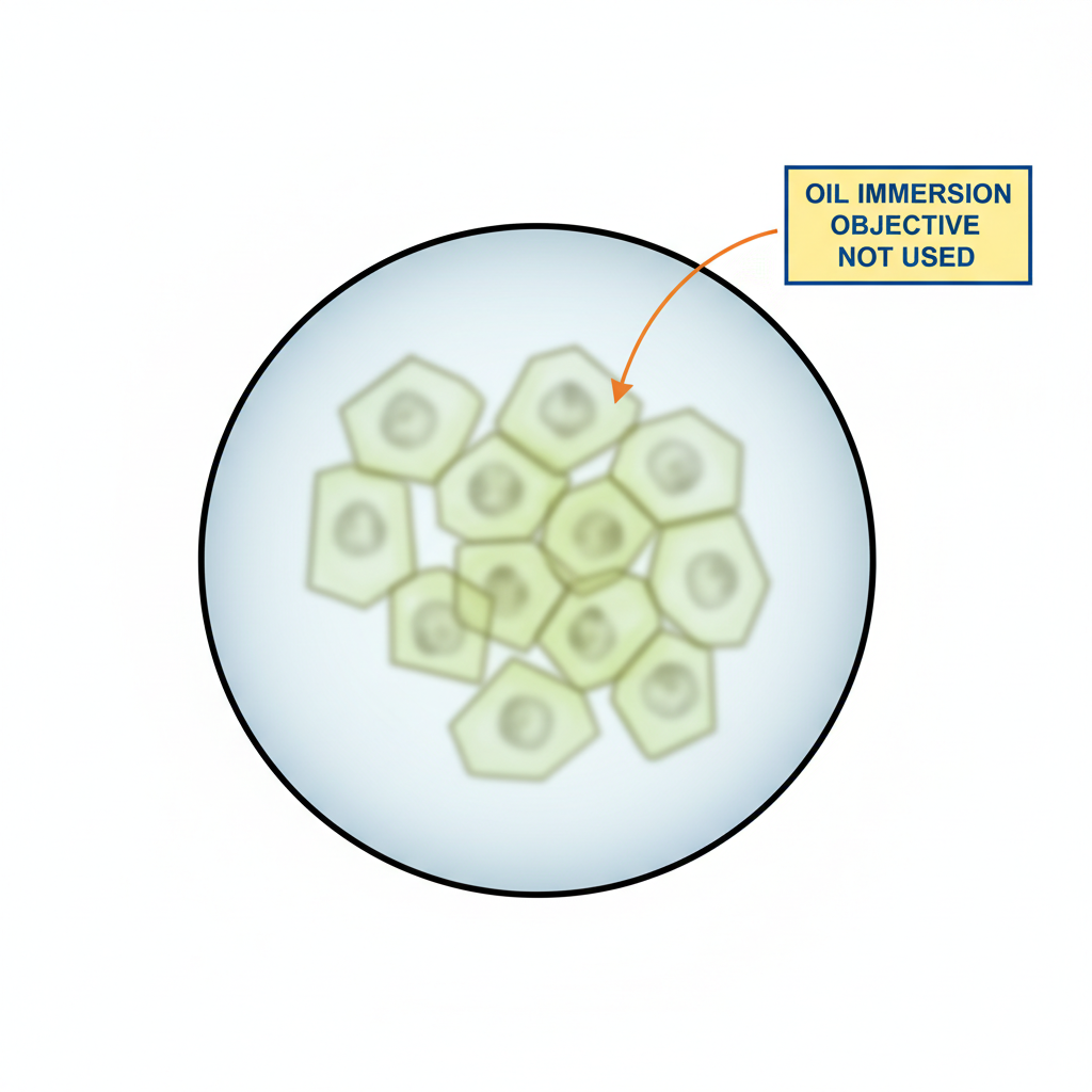
A student observes onion cells under a light microscope at high magnification. The image is blurry because the oil immersion objective has not been used. The label on the diagram reads: "Oil immersion objective not used."
a
Explain why the image appears blurry when the oil immersion objective is not used. [2]
b
Describe the correct procedure for using the oil immersion objective to obtain a clear, high-resolution image. [2]
Solutions
11QuestionParts and Functions of a Light MicroscopeConcept Practice
2 marks~3 minCriterion A
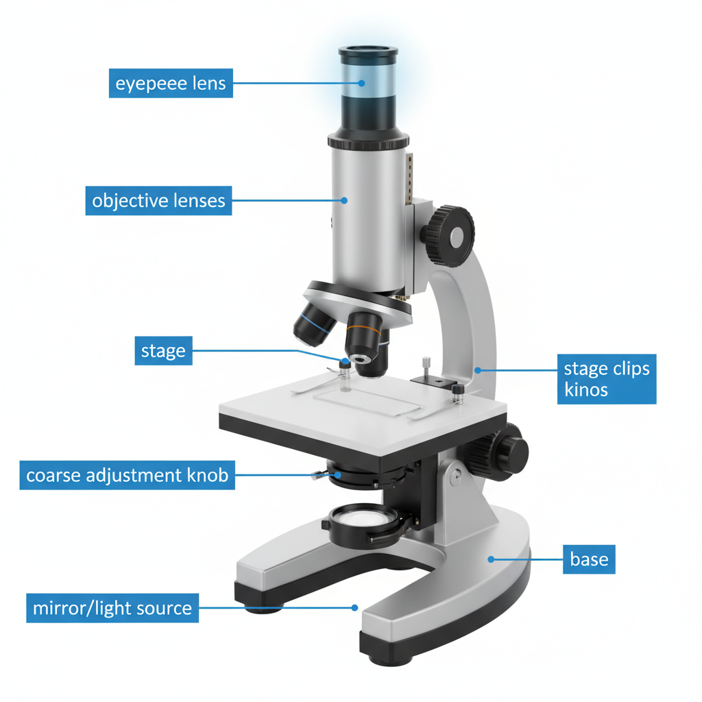
The diagram shows a light microscope with its parts labeled.
a
Describe the function of the eyepiece lens. [1]
b
A student uses the microscope. The eyepiece lens has a magnification of and the objective lens has a magnification of . Calculate the total magnification of the microscope. [1]
Solutions
12QuestionSafety and Handling of MicroscopesAssessment Practice
4 marks~6 minCriterion A
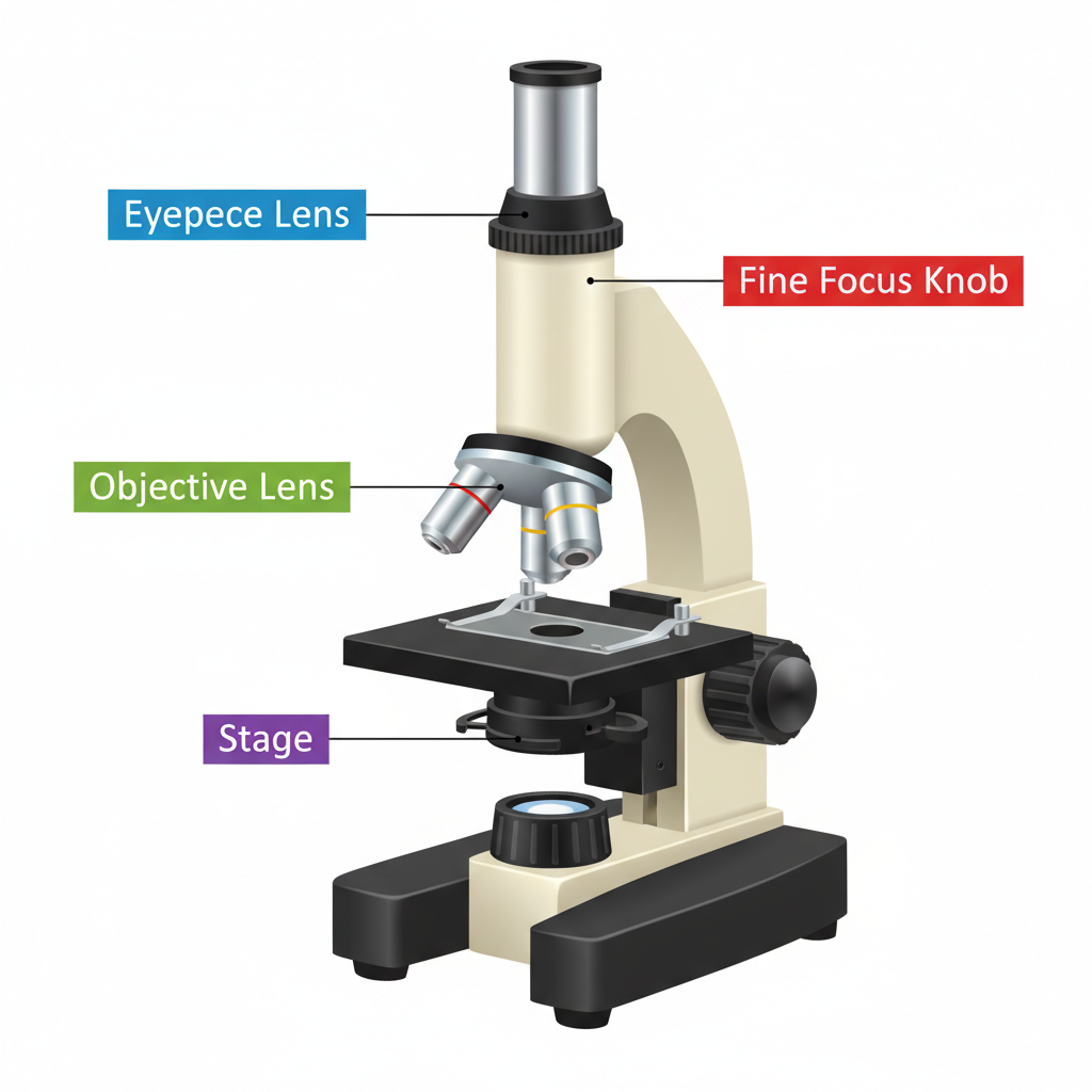
The diagram below shows a microscope with four labeled parts: eyepiece lens, objective lens, a focus knob labeled "fine focus knob," and stage.
a
Identify the labeling error in the diagram. State the correct function of each of the four labeled parts. [2]
b
Explain the correct procedure for focusing on a specimen, including the order in which the two focus knobs should be used and why this order prevents damage to the slide. [2]
Solutions
13QuestionSafety and Handling of MicroscopesConcept Practice
2 marks~3 minCriterion A
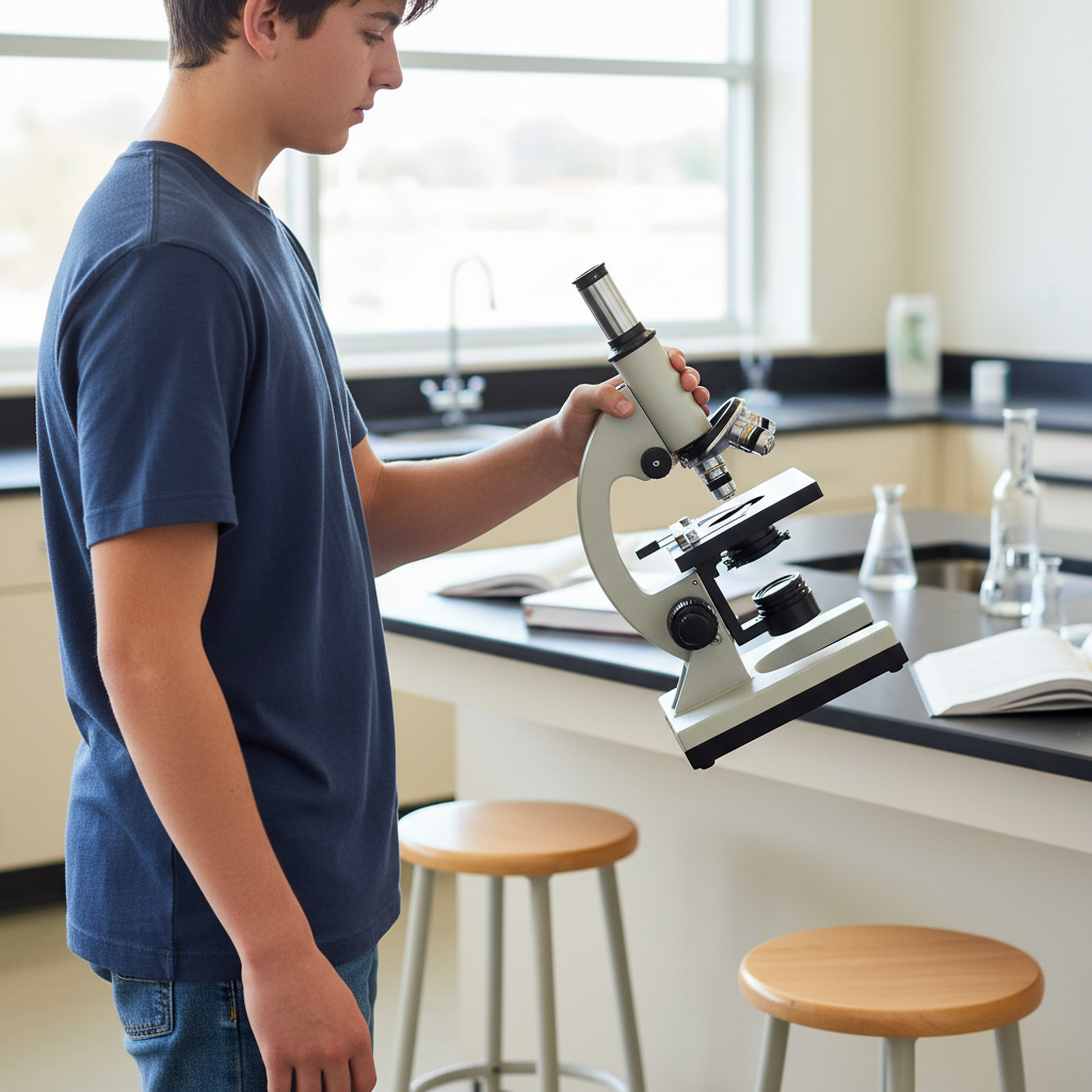
A student is seen carrying a microscope with one hand gripping the arm and the other hand swinging freely, causing the microscope to tilt dangerously.
Describe the correct two-handed technique for carrying a microscope safely. In your answer, use the terms arm, base, and stable. [2]
Describe the correct two-handed technique for carrying a microscope safely. In your answer, use the terms arm, base, and stable. [2]
Solutions
14QuestionMagnification and Field of ViewAssessment Practice
4 marks~6 minCriterion A
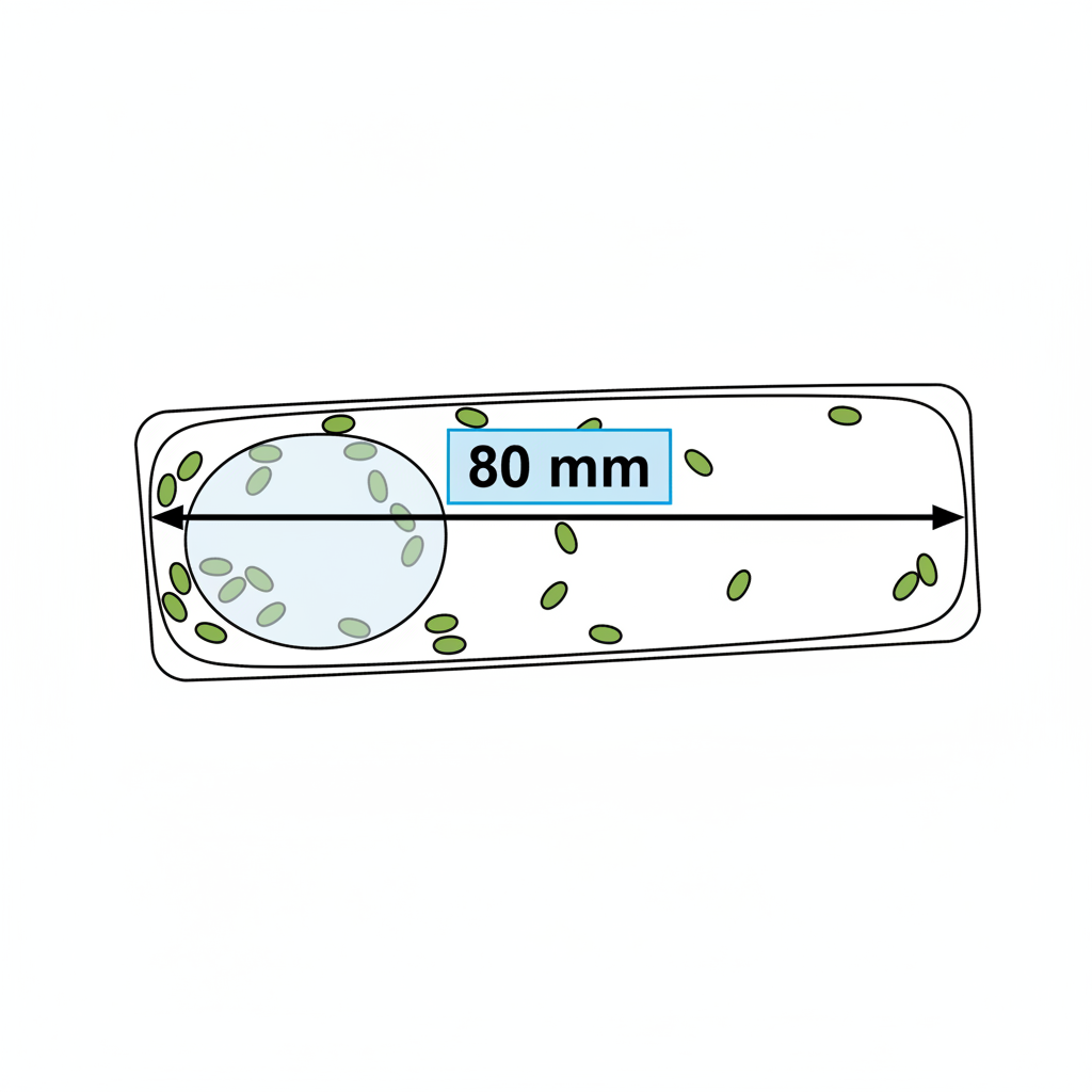
A diagram shows a plant cell viewed under a microscope at a magnification of ×400. The length of the cell in the diagram measures 80 mm.
a
Calculate the actual length of the cell. Show your working and give your answer in micrometres (µm). [2]
b
Explain why the cell appears larger under the microscope than its actual size. [1]
c
A second cell from the same plant is observed at ×100 magnification and measures 60 mm in the diagram. Explain which cell is actually longer, showing any calculation you use to support your answer. [1]
Solutions
15QuestionUsing a Microscope to View Prepared SlidesConcept Practice
2 marks~3 minCriterion A
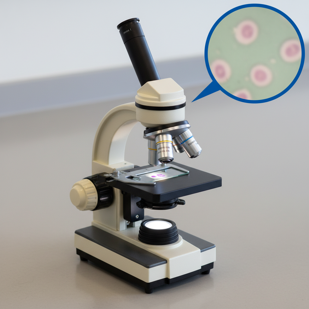
You are viewing a prepared slide of cheek cells under the low-power objective lens of a microscope. The image appears blurry.
Describe the correct sequence of steps you would take to achieve a clear focus, naming the specific microscope parts you would use.
[2]
Describe the correct sequence of steps you would take to achieve a clear focus, naming the specific microscope parts you would use.
[2]
Solutions
16QuestionParts and Functions of a Light MicroscopeAssessment Practice
4 marks~6 minCriterion D
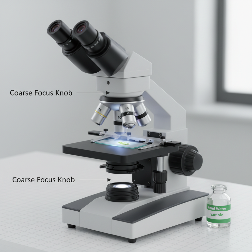
A student observing a pond water sample finds the image blurry after using the coarse focus knob, but clear after using the fine focus knob.
a
Describe one benefit of using the fine focus knob when observing living specimens. [2]
b
Explain one limitation that could occur if the fine focus knob becomes loose or misaligned during fieldwork. [2]
Solutions
17QuestionUsing a Microscope to View Prepared SlidesConcept Practice
2 marks~3 minCriterion D
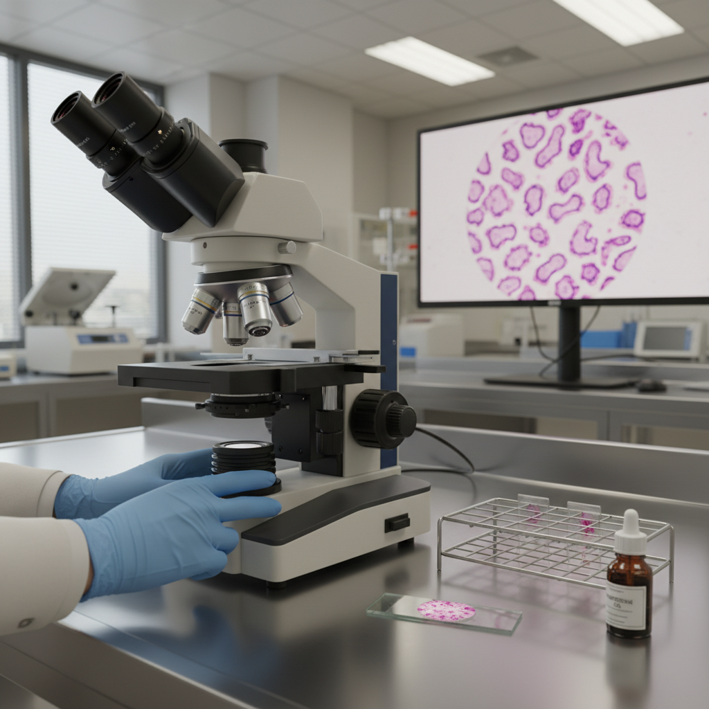
Doctors use microscopes to examine patient samples and diagnose diseases.
a
Describe one medical application of microscopy, stating what sample is examined and what doctors look for. [1]
b
Describe one limitation of using a microscope for the application you identified in part (a). [1]
Solutions
18QuestionUsing a Microscope to View Prepared SlidesAssessment Practice
4 marks~6 minCriterion C
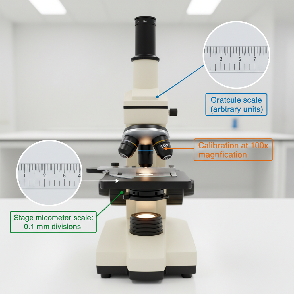
A student uses a microscope with an eyepiece graticule to measure plant cell length. At magnification, they calibrate the graticule using a stage micrometer. They then switch to magnification and measure cells using the same calibration. Five cells are measured and all give the same reading.
a
Explain why using the calibration at magnification produces inaccurate cell length measurements. [2]
b
Explain one limitation of concluding that all plant cells have the same length, based on these measurements. [2]
Solutions
19QuestionMagnification and Field of ViewConcept Practice
2 marks~3 minCriterion A
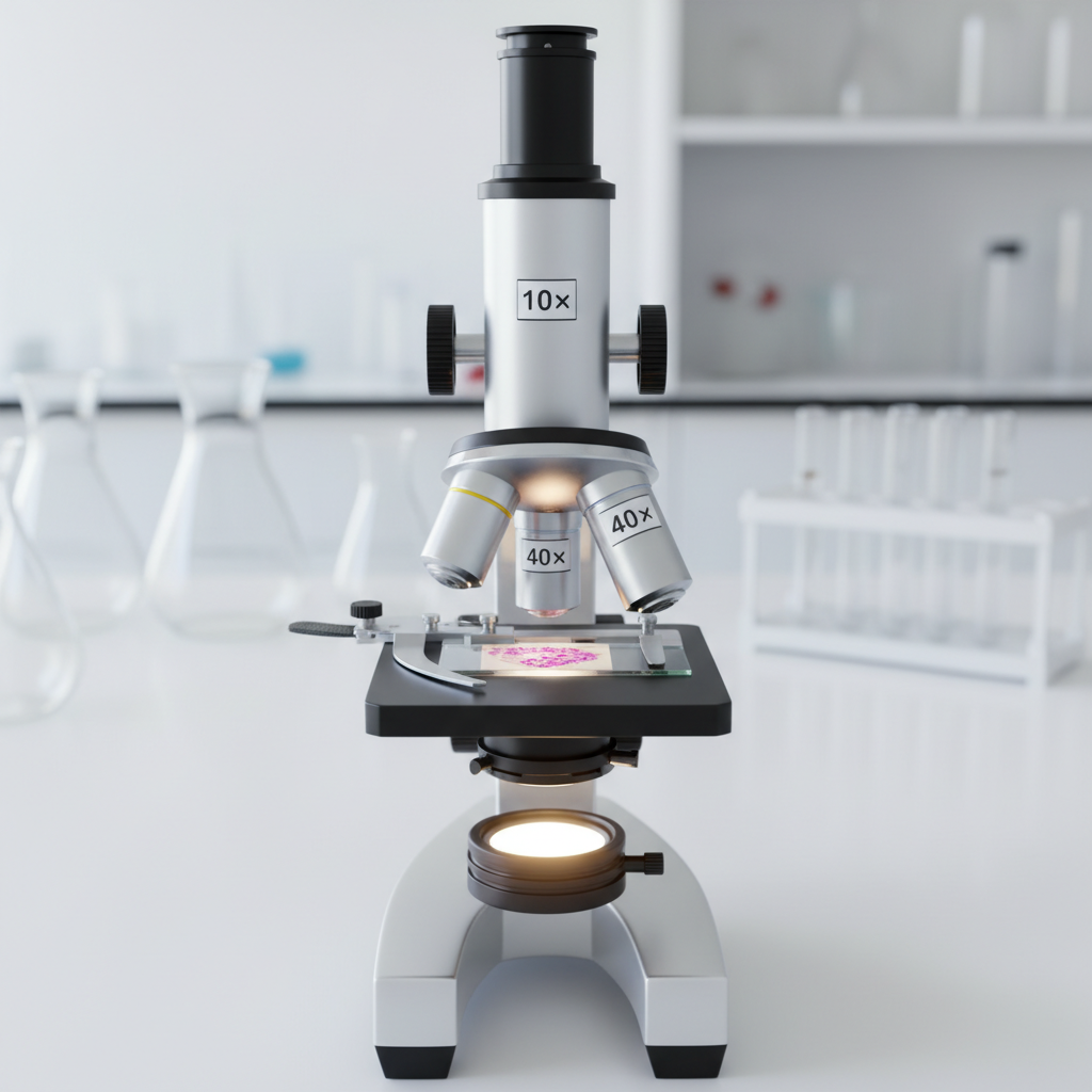
A microscope has an eyepiece lens of and an objective lens of .
(a) Calculate the total magnification of this microscope. Show the formula you use and give your answer with the correct unit. [2]
(a) Calculate the total magnification of this microscope. Show the formula you use and give your answer with the correct unit. [2]
Solutions
20QuestionParts and Functions of a Light MicroscopeAssessment Practice
5 marks~8 minCriterion C
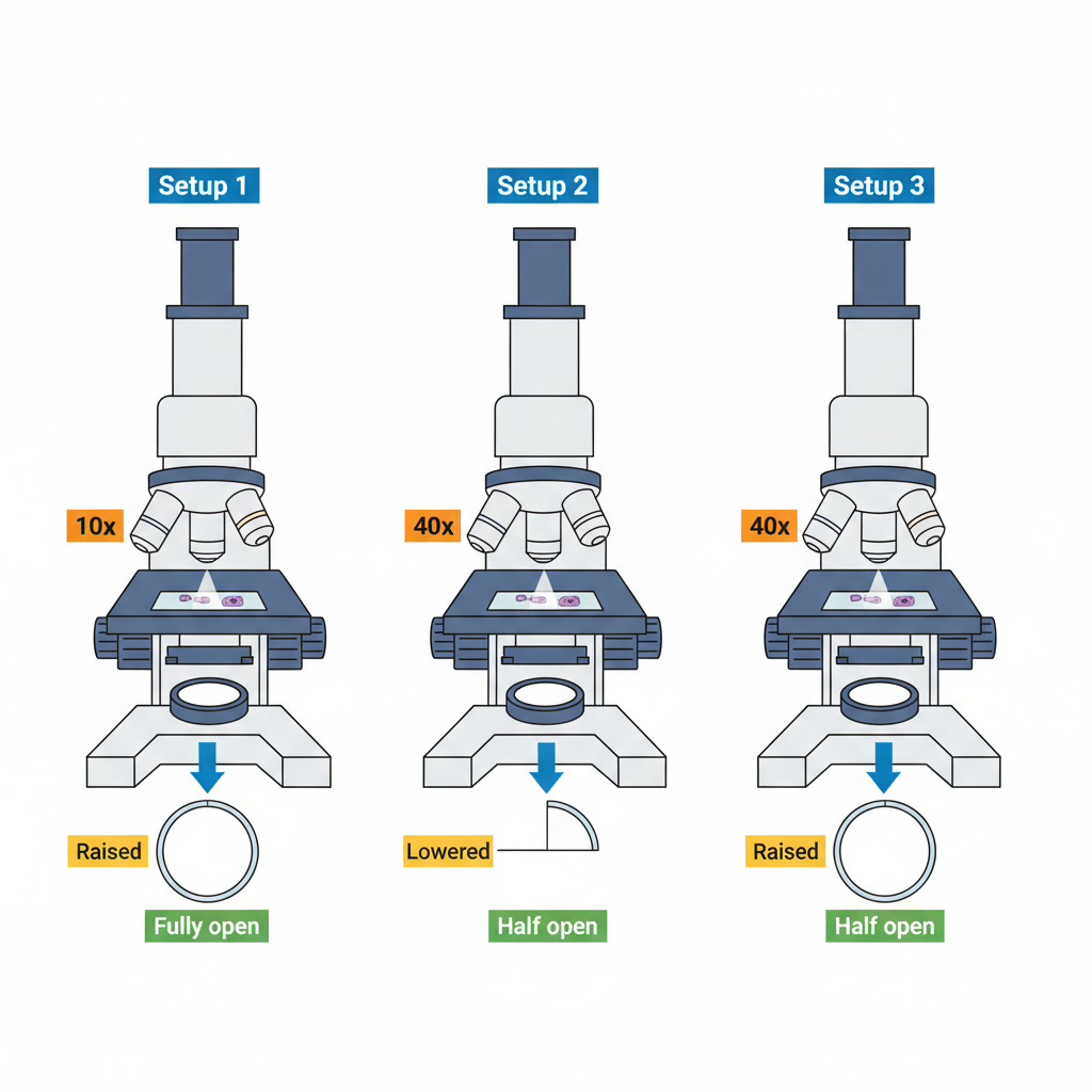
A student used a light microscope to observe onion cells under three setups.
Setup 1: objective lens , condenser raised, diaphragm fully open → cells visible but very bright, low contrast.
Setup 2: objective lens , condenser lowered, diaphragm half-open → cells clearly visible with good detail.
Setup 3: objective lens , condenser raised, diaphragm fully open → cells visible but blurry with glare.
Setup 1: objective lens , condenser raised, diaphragm fully open → cells visible but very bright, low contrast.
Setup 2: objective lens , condenser lowered, diaphragm half-open → cells clearly visible with good detail.
Setup 3: objective lens , condenser raised, diaphragm fully open → cells visible but blurry with glare.
a
State the function of the objective lens and the diaphragm in a light microscope. [2]
b
Explain how the condenser position and diaphragm setting in Setup 2 work together to improve image quality compared with Setup 1. [2]
c
Analyse the three setups and justify whether the student's claim — that Setup 2 provides the most reliable observation of onion cell structure — is valid. [1]
Solutions
Showing 20 of 79 free questions