
Biology: Differential Staining: Seeing Cell Structures Without Losing Live Cells
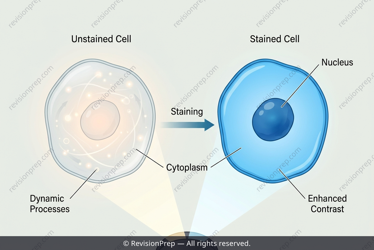
Microscopy is the art of making the invisible visible, and in cell biology, the core challenge is that most living cells are essentially transparent. This is where differential staining becomes a fundamental tool: by using dyes that bind selectively to specific cellular components, we transform a flat, ghostly image into a high-contrast snapshot where organelles stand out against the cytoplasm. The principle hinges on the chemical affinity between the stain and the target structure—for instance, methylene blue, a basic dye, binds readily to the negatively charged nucleic acids and proteins concentrated in the nucleus, rendering it a distinct dark blue mass within the otherwise pale cell. This contrast is not merely aesthetic; it is the mechanism that allows us to resolve boundaries and internal architecture that would otherwise be lost in the glare of the bright field. However, the power of staining comes with a critical trade-off. Most dyes, including methylene blue, are toxic to cells, killing them upon contact. Consequently, while a stained preparation offers exquisite structural detail, it sacrifices the very thing that makes a cell alive: its dynamic processes. The image becomes a fixed, static record—a portrait of a corpse—meaning that observations of movement, division, or active transport are impossible. Understanding this relationship between visibility and viability is essential for interpreting what a micrograph truly represents.
Start practising IB questions today
150,000+ IB-styled questions, criteria-mapped and instantly accessible.