
Biology: Epithelial Tissue Classification for IB Biology HL
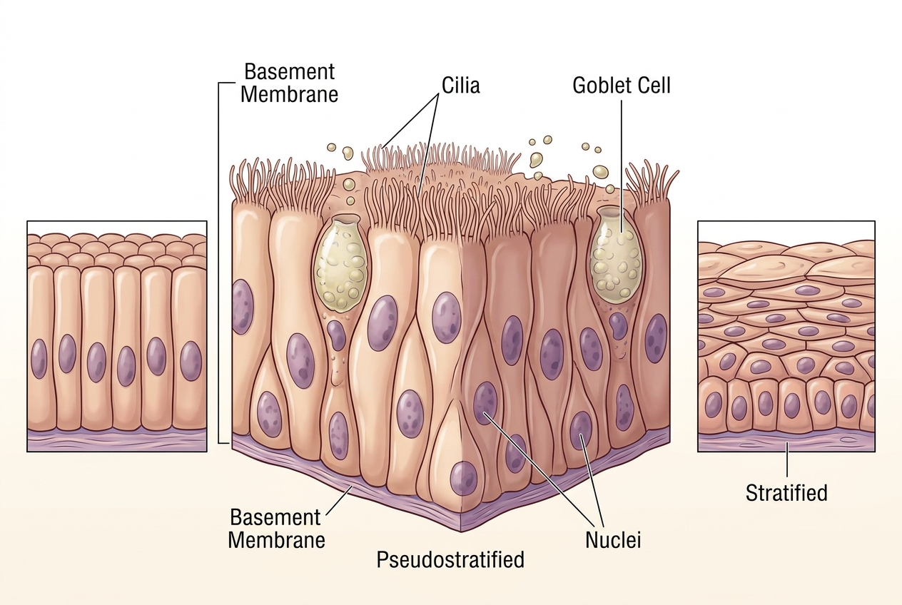
Epithelial tissue is the body’s first line of structural and functional organization—a sheet of cells that lines every surface, cavity, and tube, from blood vessels to airways. Its classification hinges on two core features: the number of cell layers (simple vs. stratified) and the shape of the surface cells (squamous, cuboidal, or columnar). Yet this simple framework hides a crucial nuance: some epithelia appear layered when they are not, and this illusion is precisely what makes histology both challenging and fascinating. The key to resolving such ambiguity lies in the basement membrane. In a truly stratified epithelium, only the basal layer touches this membrane, while upper layers are detached. However, in pseudostratified epithelium, every cell anchors to the basement membrane, even though nuclei sit at varying heights—creating a false impression of stratification. When surface cells are columnar and bear cilia, and goblet cells are interspersed for mucus secretion, the tissue is pseudostratified ciliated columnar epithelium. This arrangement maximizes surface area for particle trapping and clearance, as seen in the trachea, where the coordinated beating of cilia propels mucus upward. Understanding this distinction—between true layers and nuclear positioning—is essential for interpreting any epithelial section correctly.
Start practising IB questions today
150,000+ IB-styled questions, criteria-mapped and instantly accessible.