
Biology: Rethinking the Fluid Mosaic Model
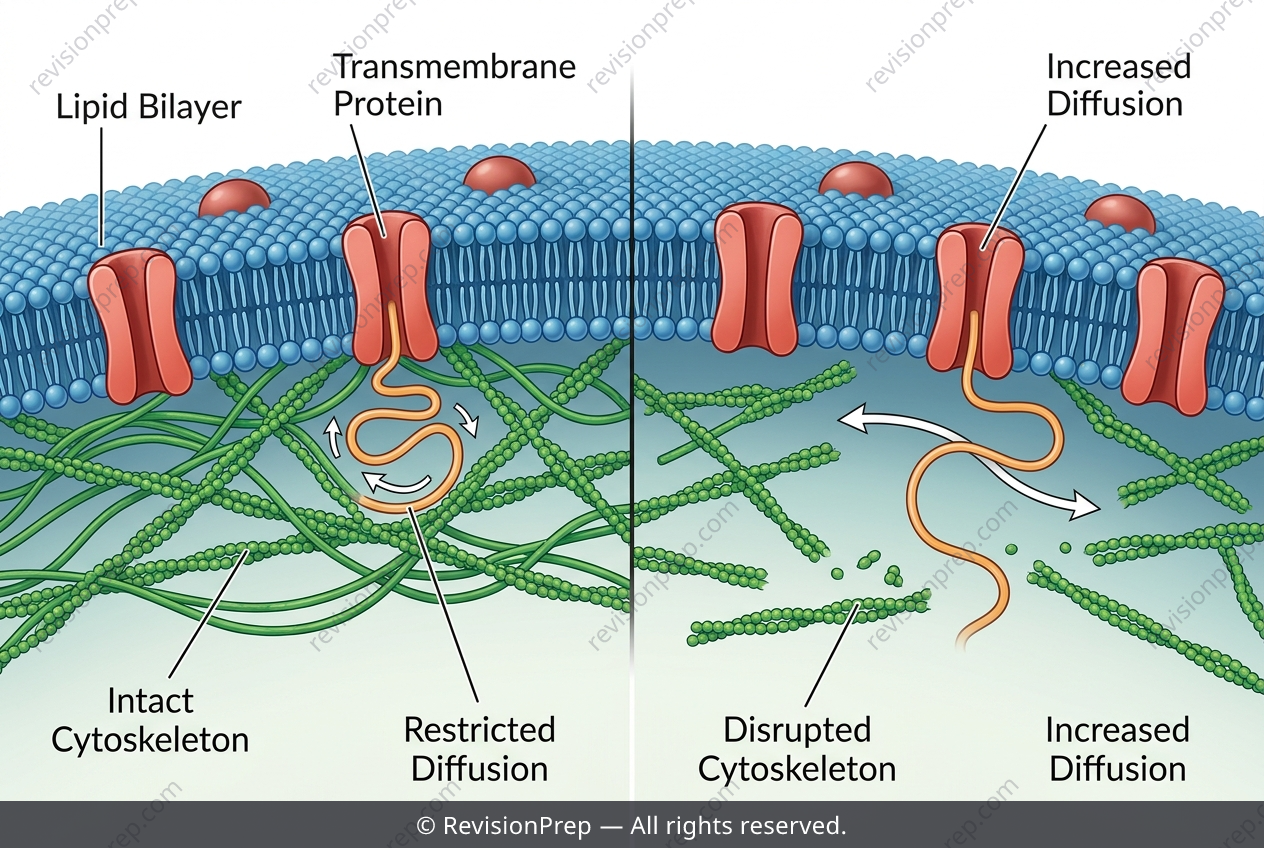
The plasma membrane is far more than a passive barrier—it is a dynamic, crowded stage where protein mobility dictates cellular function. At the heart of this lies the concept of membrane fluidity, often quantified by the lateral diffusion coefficient D, which describes how quickly proteins like glycophorin move within the lipid bilayer. Using single-particle tracking, researchers measure the mean squared displacement (MSD) over a short time interval, applying the relationship MSD = 4DΔt to extract D. This seemingly simple equation unlocks a deeper story: the membrane is not a uniform sea of lipids, but a patchwork constrained by an underlying cytoskeletal meshwork. In human erythrocytes, actin filaments form a structural scaffold beneath the membrane, tethering transmembrane proteins via linker proteins like spectrin and ankyrin, and creating “corrals” that limit free diffusion. When this meshwork is disrupted—for instance, by depolymerising actin—the measured D increases dramatically. This observation directly challenges the classic fluid mosaic model, which predicted unrestricted protein movement. Understanding this interplay between lipid fluidity, protein anchoring, and cytoskeletal barriers is essential for grasping how cells regulate signalling, adhesion, and membrane organisation in health and disease.
Start practising IB questions today
150,000+ IB-styled questions, criteria-mapped and instantly accessible.