
Physics: From Alpha Scattering to X-Ray Imaging
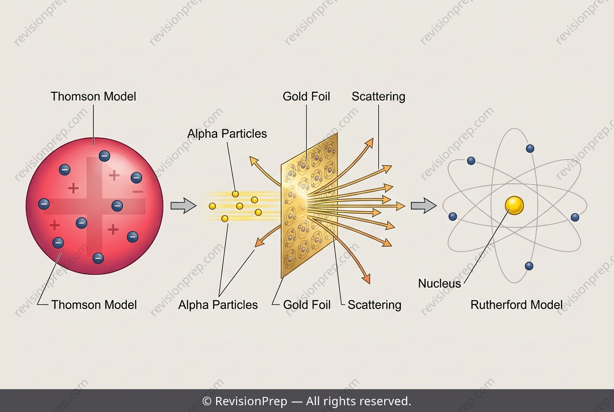
How do we know what an atom looks like if we can’t see it? The answer lies in a chain of empirical refinements—each experiment forcing scientists to revise their mental picture. Thomson’s plum pudding model imagined a diffuse, positively charged sphere with electrons scattered like raisins. But Rutherford’s gold foil experiment shattered this view: firing alpha particles at a thin foil, he observed that while most passed straight through, a few rebounded at angles greater than 90°. Such large-angle deflections are impossible under Thomson’s model, because a spread-out positive charge exerts only weak, scattered repulsive forces—nowhere near concentrated enough to fling a fast alpha particle backward. This single anomaly triggered a paradigm shift. Rutherford replaced the pudding with a nuclear model: a tiny, dense, positively charged nucleus surrounded by mostly empty space, with electrons orbiting far away. The relationship between charge concentration and deflection (essentially Coulomb’s law, F = kq₁q₂/r²) became the key mechanism—the tighter the positive charge, the stronger the repulsive force at close range. Crucially, this refined understanding of atomic structure didn’t just satisfy curiosity; it gave physicists a precise map of how matter interacts with radiation. By knowing where the nucleus sits and how electrons absorb energy, scientists could calculate and control X-ray penetration and dosage—minimising tissue damage while maximising imaging clarity. Thus, a model born from scattered particles now underpins safer medical diagnostics, linking fundamental physics directly to patient care.
Start practising IB questions today
150,000+ IB-styled questions, criteria-mapped and instantly accessible.