
Physics: Nuclear Decay, Mass & Medical Imaging
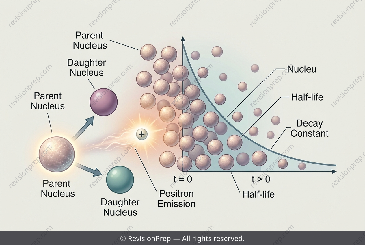
Radioactive decay kinetics and mass-energy equivalence are two pillars of nuclear physics that come together beautifully in medical imaging. At its heart, this topic asks: how do unstable nuclei transform, and where does the released energy actually come from? For fluorine-18, a positron emitter used in PET scans, the decay follows an exponential law where the activity A drops as A = A₀e^(−λt), with the decay constant λ linked to the half-life by λ = ln2 / T₁/₂. This relationship lets you predict how many radioactive atoms remain at any moment, which is crucial for timing a scan or calculating a safe dose. But the deeper story lies in the energy released during the decay. When a proton in fluorine-18 becomes a neutron, a positron is ejected—yet the mass difference between parent and daughter atoms does not directly give the energy. Because atomic masses include electrons, and positron emission creates an extra particle, you must account for two electron masses in the mass defect. Only then does E = Δmc² reveal the true Q-value, the energy carried away by the positron and the daughter nucleus. This connection between measurable masses and the energy of nuclear transitions is what powers everything from cancer imaging to understanding stellar fusion.
Start practising IB questions today
150,000+ IB-styled questions, criteria-mapped and instantly accessible.