
Physics: Radioactive Decay, Counts & PET Imaging
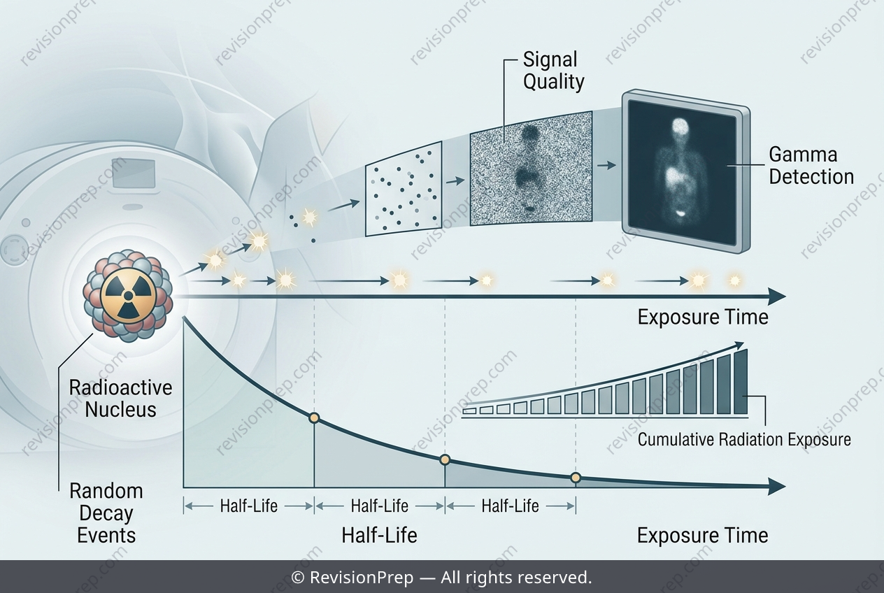
Radioactive decay is the spontaneous, random transformation of an unstable nucleus into a more stable one, a process that lies at the heart of nuclear physics and modern medical imaging. In positron emission tomography (PET), a tracer with a half-life of about 110 minutes emits positrons that annihilate with electrons, producing gamma rays detected by the scanner. This randomness is not a flaw—it is the very property that makes the technique safe and practical. Because decay is spontaneous, the tracer’s activity falls exponentially over time, following the relationship A = A₀(1/2)^(t/T₁/₂). After several half-lives, the activity becomes negligible, meaning the patient’s radiation dose is self-limiting, unlike a stable substance that would irradiate indefinitely. Yet the same randomness introduces statistical fluctuations in the number of detected events. A short scan window yields too few counts, producing a noisy, low-resolution image, while a long window allows tracer decay to reduce the count rate and patient movement to blur the result. The radiographer must balance these competing effects, choosing a scan duration that accumulates enough gamma-ray events for clarity without extending into the regime where decay and motion degrade the image. This interplay between decay kinetics, counting statistics, and imaging quality defines the core challenge of PET optimization.
Start practising IB questions today
150,000+ IB-styled questions, criteria-mapped and instantly accessible.