
Physics: Radioactive Isotopes in Medical Imaging
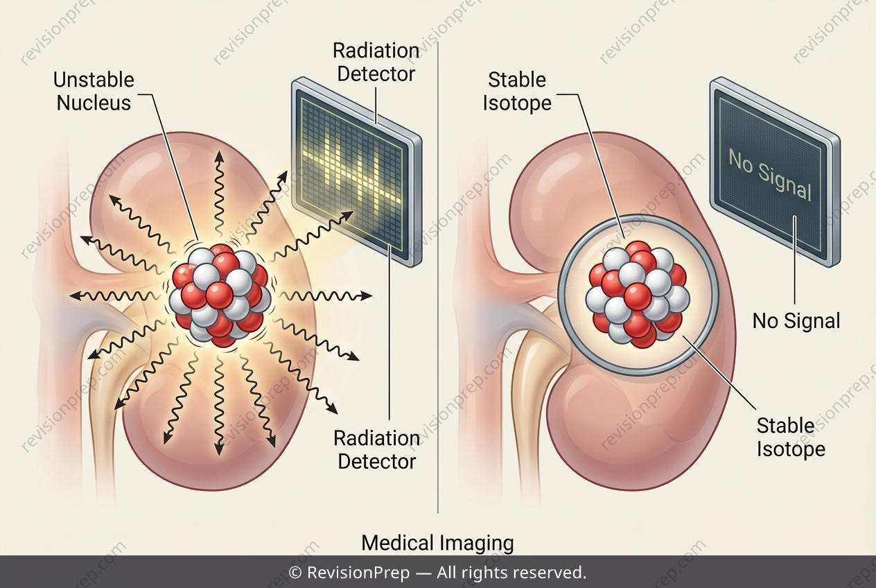
Radioactive decay isn’t just a theoretical curiosity—it’s the engine behind some of modern medicine’s most precise diagnostic tools. At the heart of this lies the concept of isotope properties: while all isotopes of an element share the same number of protons, they differ in neutron count, which dictates stability and decay behaviour. Technetium-99m, a metastable isotope, exemplifies this perfectly. Its nucleus releases excess energy as gamma radiation—high-energy photons that can pass through body tissue—rather than as heavier particles, making it uniquely suited for external detection. This is where the connection between decay type, half-life, and medical imaging becomes clear. A stable isotope of technetium, by definition, emits no radiation, so it would be invisible to a gamma camera and useless for imaging. In contrast, the gamma rays from 99mTc penetrate tissue and are captured externally, producing a functional image of organs like bones or the heart. Crucially, its half-life of roughly 6 hours balances two competing needs: long enough to perform the scan, yet short enough to minimise the patient’s radiation dose. This interplay—between emission type, penetration, and decay timing—is what transforms a nuclear physics concept into a life-saving clinical procedure.
Start practising IB questions today
150,000+ IB-styled questions, criteria-mapped and instantly accessible.