
Form and Function
The AHL logic linking structure to function — from sliding filaments to stem cell fate
Quick facts
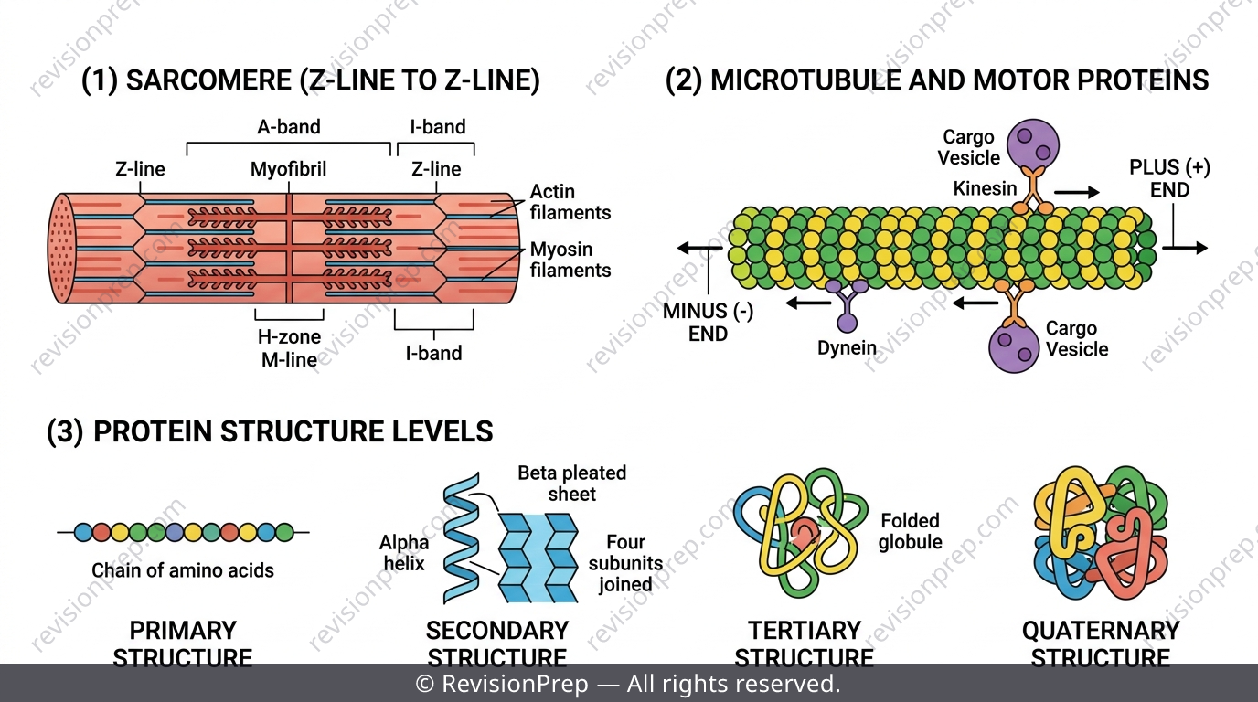
IB Biology's Form and Function theme keeps asking the same question in new disguises: given this structure, what function must follow? Nowhere is that clearer than in Muscle and Motility, an AHL-exclusive subtopic where sliding filaments, motor proteins, and cardiac conduction all show up on HL Paper 1 and Paper 2. Add cell specialisation, stem cell potency, and the four levels of protein structure, and you've got a theme that examiners love to test with 'explain the mechanism' questions. This teaser covers the five ideas that generate the most lost marks — the ATP logic of the cross-bridge cycle, kinesin versus dynein direction, the AV node delay, potency hierarchy, and protein folding — so you can walk into Paper 2 ready to reason through unfamiliar structure-function scenarios instead of memorising isolated facts. The full revision notes go deeper into every example question and trap.
What you’ll be able to do
The Sliding Filament Model & Cross-Bridge Cycle
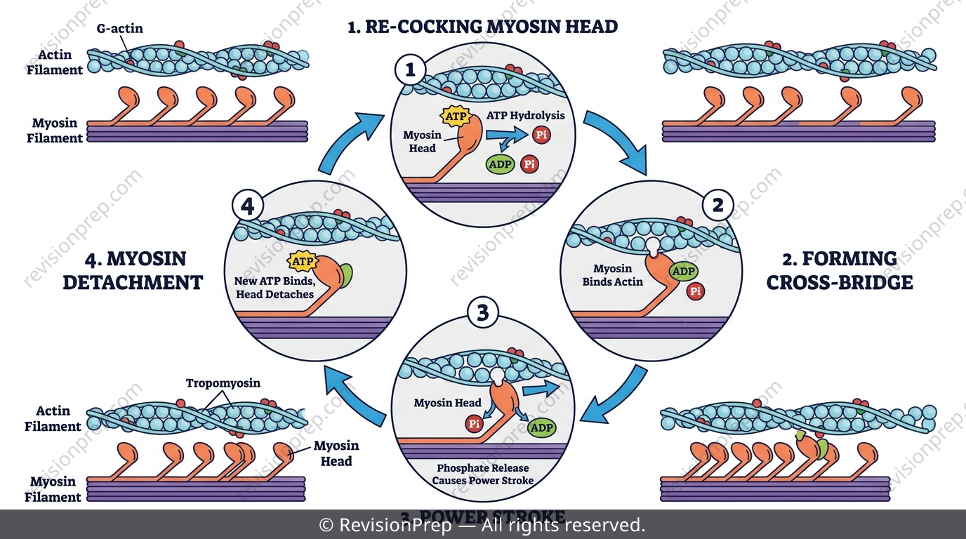
Skeletal muscle contracts because thick myosin filaments and thin actin filaments interdigitate inside a sarcomere and slide past each other — the filaments themselves never shorten. An action potential triggers Ca2+ release from the sarcoplasmic reticulum, and Ca2+ binds troponin, causing tropomyosin to shift and expose myosin-binding sites on actin. The cross-bridge cycle then runs in a strict order: ATP hydrolysis re-cocks the myosin head, the head binds actin, phosphate release drives the power stroke, ADP leaves, and only fresh ATP binding causes detachment.
Exam tip
Reverse the common logic: ATP is needed to BREAK cross-bridges (detachment), not to form them. Write it that way and you'll nail the 'why can't the muscle relax' question.
Common mistake
Never say filaments 'shorten' during contraction — IB mark schemes penalise this. The A-band stays constant (thick filament length is fixed); only the I-band and H-zone narrow as thin filaments slide inward.
Mini summary
Ca2+ binds troponin (not tropomyosin); ATP hydrolysis cocks the head, ATP binding detaches it; filaments slide, they don't shorten.
Motor Proteins: Kinesin, Dynein & Microtubule Polarity
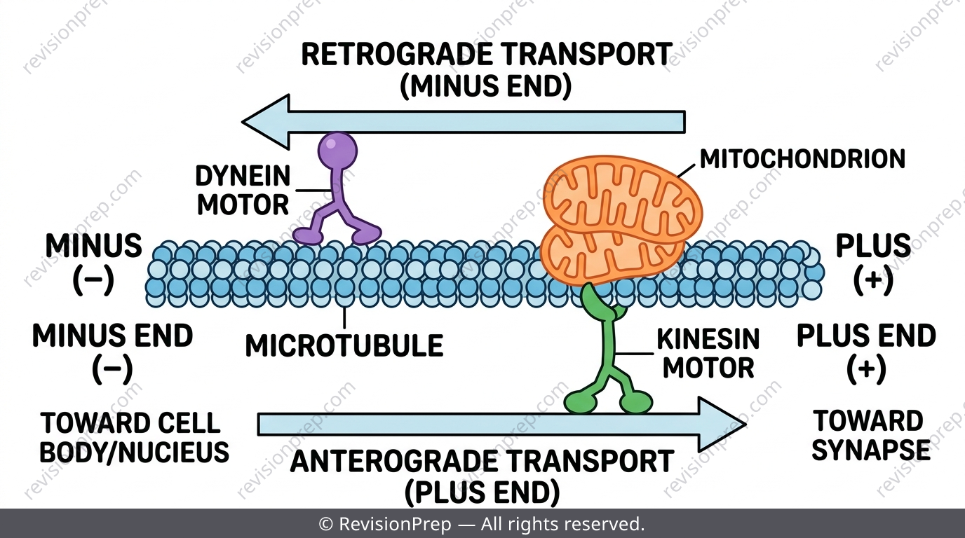
Cells move cargo along two cytoskeletal tracks: microtubules (walked by kinesin and dynein) and actin microfilaments (walked by myosin). Microtubules have polarity — a plus end, usually toward the cell periphery or synapse, and a minus end toward the cell centre. Kinesin is plus-end-directed while dynein is minus-end-directed, and many organelles carry both motors simultaneously, so blocking one can unmask the other's pull in the opposite direction.
Exam tip
Match the motor to the direction stated in the stem, not the other way round — kinesin = plus end, dynein = minus end, every time.
Common mistake
Don't assume blocking one motor stops an organelle dead. If a mitochondrion carries both kinesin and dynein, inhibiting kinesin can cause it to reverse direction as dynein takes over.
Mini summary
Kinesin walks to the plus end; dynein walks to the minus end — direction is set by the motor, not the cargo.
Cardiac Muscle & the Conduction System
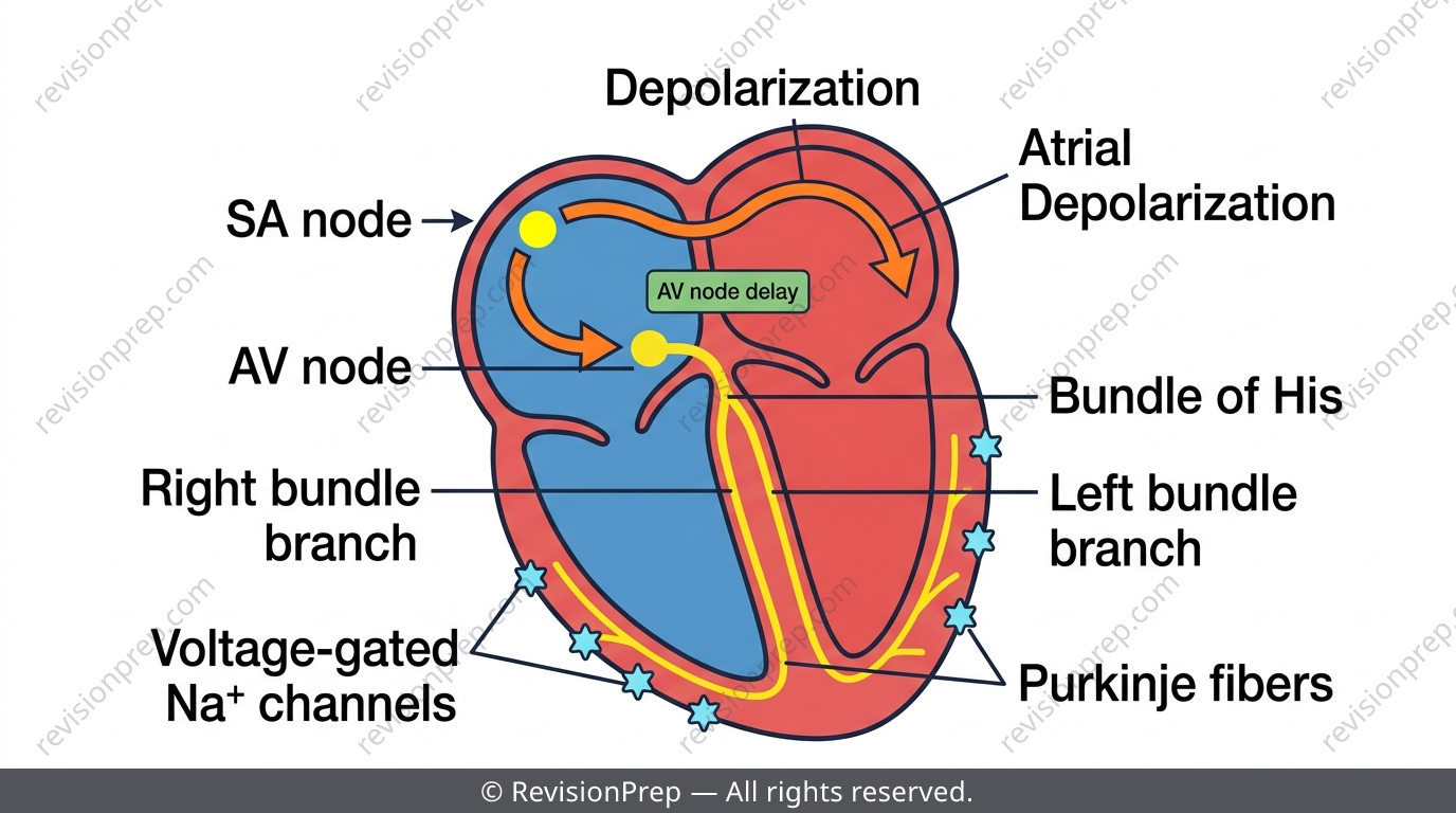
Cardiac muscle contracts under its own built-in rhythm: the SA node generates spontaneous depolarisation, and voltage-gated Na+ channels drive the fast action potential upstroke as it spreads through atrial and ventricular muscle. A conduction delay at the AV node lets the atria finish contracting before the ventricles begin, keeping the pumping sequence coordinated.
Exam tip
Voltage-gated Na+ channels control the speed of impulse propagation through working muscle — blocking them slows or desynchronises conduction. They do NOT raise heart rate; that's a pacemaker Ca2+-channel effect at the SA node.
Common mistake
Naming the wrong channel's role loses the mark even if 'slower conduction' is otherwise correct — keep Na+ channels (propagation speed) separate from SA node pacemaker channels (rate).
Mini summary
SA node sets the rhythm, Na+ channels drive propagation speed, and the AV node delay sequences atrial-then-ventricular contraction.
Cell Specialisation & Stem Cell Potency
Every somatic cell keeps (almost) the same genome, but differentiation switches genes on and off via differential gene expression — not gene loss. Potency describes how many fates a stem cell can still choose: totipotent cells can become any cell type including extra-embryonic tissue, pluripotent cells can become any body cell type but not extra-embryonic tissue, and multipotent cells are restricted to one lineage. Specialisation often means discarding structures too, like mature red blood cells losing their nucleus and organelles to maximise room for haemoglobin.
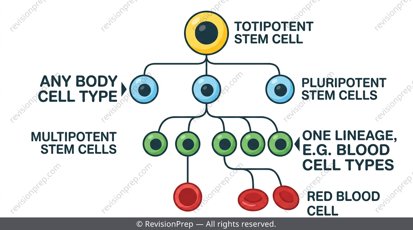
| Potency | Range of cell fates |
|---|---|
| Totipotent | Any cell type, including extra-embryonic tissue |
| Pluripotent | Any body cell type, but not extra-embryonic tissue |
| Multipotent | One lineage only (e.g. blood stem cells → blood cells) |
Exam tip
Always link a discarded structure to the freed-up function it enables — losing the nucleus isn't just 'less stuff', it's more space for gas exchange.
Common mistake
Don't imply cells lose DNA during differentiation. Every somatic cell retains the full genome; only gene expression changes.
Mini summary
Potency narrows: totipotent > pluripotent > multipotent — driven by gene expression changes, not DNA loss.
Levels of Protein Structure
Proteins are chains of 20 amino acids joined by peptide bonds; the sequence (primary structure) determines every fold above it, which is why a single amino acid substitution can wreck function, as in sickle-cell haemoglobin. Secondary structure is regular hydrogen-bonded patterns (alpha helix, beta pleated sheet); tertiary structure is the overall 3D fold of one polypeptide held by R-group interactions including hydrogen bonds, ionic bonds, disulfide bridges, and hydrophobic interactions; quaternary structure assembles multiple polypeptide subunits together, as in haemoglobin's four subunits.
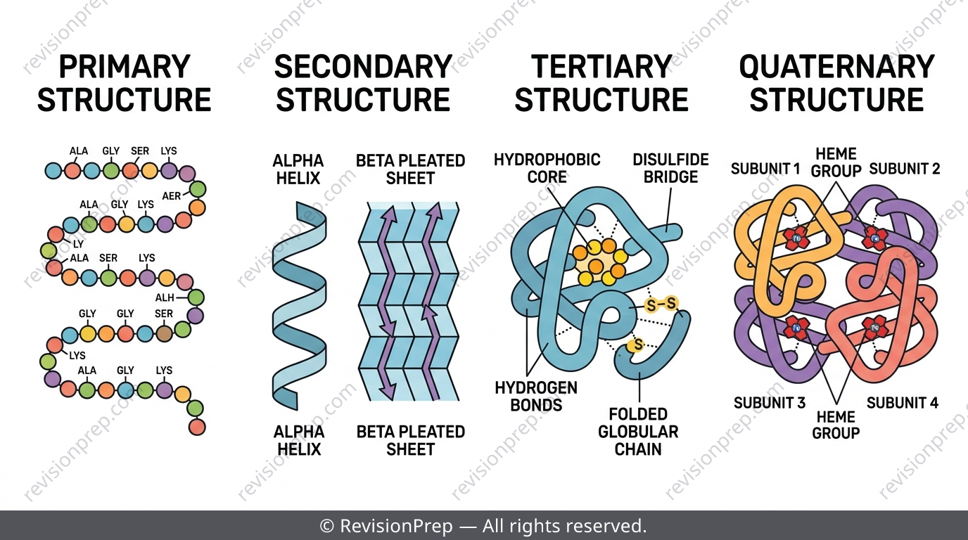
| Level | Description | Key bonds/interactions |
|---|---|---|
| Primary | Sequence of amino acids | Peptide bonds |
| Secondary | Repeating local folding pattern | Hydrogen bonds along backbone |
| Tertiary | Overall 3D fold of one polypeptide | H-bonds, ionic bonds, disulfide bridges, hydrophobic interactions |
| Quaternary | Assembly of multiple polypeptide subunits | Same R-group interactions between subunits |
Exam tip
If a question mentions a single amino acid change affecting the whole protein, trace it upward: primary → tertiary fold → quaternary assembly → function lost.
Mini summary
Primary sequence dictates secondary, tertiary, and quaternary folding — one amino acid change can cascade through all four levels.
Quick formula sheet
Practice questions
- Name the ion that binds troponin to trigger the exposure of myosin-binding sites on actin.
- State whether kinesin travels toward the plus end or the minus end of a microtubule.
- Define a totipotent stem cell.
- Explain why the A-band width stays constant during contraction while the I-band narrows.
- Explain the role of the AV node delay in coordinating atrial and ventricular contraction.
- Describe how differential gene expression, rather than gene loss, explains cell specialisation.
- A muscle fibre is treated with a chemical that blocks ATP binding to myosin heads. Predict and explain the effect on relaxation.
- An antibody blocks kinesin's motor domain in a neuron where a mitochondrion carries both kinesin and dynein. Predict the immediate change in the mitochondrion's movement.
- Explain why a single amino acid substitution in haemoglobin can disrupt its quaternary structure and function.
Frequently asked questions
What triggers muscle contraction at the molecular level?+
An action potential causes Ca2+ release from the sarcoplasmic reticulum. Ca2+ binds troponin, which shifts tropomyosin to expose myosin-binding sites on actin, allowing the cross-bridge cycle to begin.
What is the difference between kinesin and dynein?+
Both are motor proteins that walk along microtubules, but kinesin moves toward the plus end (usually the cell periphery) while dynein moves toward the minus end (usually the cell centre).
Why can't a muscle relax without ATP if ATP is used to contract?+
ATP hydrolysis re-cocks the myosin head for the next power stroke, but it's fresh ATP BINDING to the head that causes it to detach from actin. Without ATP, heads stay attached and the muscle can't relax — this is why rigor mortis occurs.
Is Muscle and Motility examined at IB Biology SL?+
No — Muscle and Motility, along with enzyme kinetics and endosymbiotic theory, is AHL-exclusive and only appears on HL papers.
What is the difference between totipotent, pluripotent, and multipotent stem cells?+
Totipotent cells can form any cell type including extra-embryonic tissue, pluripotent cells can form any body cell type but not extra-embryonic tissue, and multipotent cells are restricted to a single lineage.
Do muscle filaments actually shorten during contraction?+
No. Thick and thin filaments slide past each other — their individual lengths never change. IB mark schemes specifically penalise answers that describe filaments as 'shortening'.
Get the full Form and Function revision notes
Related articles